-->

Monday, December 23, 2013

The Link Between Autoimmunity and Infectious Disease


Autoimmunity occurs when the immune system begins to wrongly attack self-antigens or various parts of the body it’s responsible for protecting. There are mechanisms in place to prevent this kind of problem, while still allowing B and T-cells the variability needed to combat a wide array of pathogens (disease causing agents). Mechanisms are in place during the production of B and T-cells that check them for auto reactivity, such as central and peripheral tolerance. The breakdown of these tolerance mechanisms causes autoimmunity. When central tolerance mechanisms breakdown, auto reactive B-cells escape negative selection, in the form of apoptosis (cell death), and are released into the lymphatic system. It is when these auto reactive B-cells become aberrantly activated through things like B and T-cell discordance or T-cell bypass that an attack on host tissues begins.
            Some autoimmune diseases are caused by the production of autoantibodies against self-molecules, which are produced when auto reactive B and T-cells escape tolerance mechanisms. The production of autoantibodies against key components in the immune system, like inflammatory cytokines (signaling molecules) can negatively affect the ability of the immune system to effectively clear pathogens and prevent disease. In other words, autoimmunity can predispose people to infection by various pathogens, which they would normally be able to eradicate if their immune system was uncompromised.
            One of the most vital aspects of the immune system is the ability of the various immune cells to effectively communicate with one another and influence the environment around them. This is done through the use of signaling molecules known as cytokines. They are a diverse group of soluble proteins, peptides or glycoproteins that are secreted by specific immune cells in order to elicit a specific response. Therefore many times when an infection is detected inflammatory cytokines such as TNF-α are released, which induces things like vasodilation. Thus, by increasing the amount of immune cells flowing to the site of infection the environment around the pathogen is effectively skewed to favor the immune system. Cytokines can also act as effector molecules to polarize T-cell responses in order to rid the body of the pathogen in the safest and most effective way possible. For instance, if an extracellular pathogen like Streptococcus (the bacteria that causes strep throat) began to infect your nasal system you would want a Th2 (antibody mediated) response to be deployed as opposed to a Th1 (CTL mediated) response. A Th2 response is much safer and more effective against extracellular pathogens because, unlike a Th1 response, a Th2 antibody-mediated response doesn’t involve the release of cytotoxic granules or pro-inflammatory molecules, which could really damage these sensitive tissues. In order for a T-cell response to be skewed towards a Th2 response the cytokine IL-4 must be present and continue to remain in the environment. So, if a person suffered from an autoimmune disease, which produced autoantibodies against IL-4 they would be unable to effectively combat extracellular pathogens like Streptococcus, and in a sense they would be classified as immune compromised.

Sunday, December 22, 2013

SCARF1: A Novel Receptor for Apoptotic Clearance

            Autoimmunity is one of the most difficult to treat and unfathomable types of disease, because we are not fighting a virus, bacterium, or parasite: we are fighting our own body. Usually, doctors can count on the immune system to help them out when their patients are sick; it is the job of this system to ensure that any foreign threat to the body is eliminated. But the nature of autoimmunity is such that the biggest ally we have in the quest to keep ourselves healthy turns against us and begins attacking that which it has evolved to protect.
There are many classes of autoimmune disease, each with a very different cause. The immune system is so diverse and complicated that a mutation in one of its parts can affect the entire system and ultimately manifest in disease. There are myriad ways in which this can happen, but the result is what is called “breaking tolerance”. When tolerance is broken, the immune system recognizes some small component of the body as a foreign object and mounts an immune response against it. This can cause differing amounts of damage, depending on how prevalent the component is, and if its recognition and destruction leads to the labeling of more self proteins as targets. There is the danger of a phenomenon called epitope spreading, which happens when a cell is targeted and destroyed, releasing its contents into the body. The immune system has not been desensitized, or “tolerized” to the proteins inside a cell, as it should have no need to recognize them in a healthy body. When this happens in the context of an already active self-targeted immune response, immune cells may further target the otherwise normal contents of the dying cells, leading to a more serious attack throughout the body.
            One important safeguard against autoimmunity is the safe breakdown clearance of apoptotic cells. An apoptotic cell is one that is infected, compromised, or simply too old and losing effective function. These cells are marked for uptake by phagocytes, which are a class of cell types that uptake and destroy their targets, breaking down anything in the cell that could be toxic if released into the body. It is known how and when phagocytes such as macrophages and dendritic cells destroy their targets, but the specifics of their identification are little investigated. In their paper “The scavenger receptor SCARF1 mediates the clearance of apoptotic cells and prevents autoimmunity”, Zaida G. Ramirez-Ortiz et al identify and characterize the receptor SCARF1 which, allows phagocytes to recognize their targets for destruction. SCARF1 is a transmembrane protein which has homologs even in the simple research model C. elegans, and acts by binding to a C1q and phosphatidylserine complex. Phosphatidylserine is a part of the inside of the cell membrane, and becomes exposed on the exterior portion of the membrane only when the cell needs to be phagocytosed. SCARF1, the researchers found, cannot recognize and destroy cells without this component bound to C1q, a peptide which also plays a role in the complement system. High concentrations of this peptide near a cell marked with phosphatidylserine cause a complex to form, which binds to SCARF1 and results in successful phagocytosis. 
   
Cover image expansion
1. Macrophage Engulfing Apoptotic Cells


Friday, December 20, 2013

A New Vaccine to Protect Against Malaria


Malaria mosquito (4)
Malaria protozoan (5)
A New Vaccine to Protect 
Against Malaria


Malaria cases by country (6)
Malaria is a serious mosquito-borne illness that is present in tropical and sub-tropical countries all over the world. Roughly 3.3 billion people, almost half of the world’s population, live in areas where malaria is endemic (1). Malaria is caused by a unicellular eukaryotic organism called a protozoan. The protozoan that causes malaria, Plasmodium falciparum (Pf), replicates in human liver and red blood cells causing these cells to die. Uninfected mosquitos can then pick up the sporozoite by biting an infected person, thus propagating infection. Malaria causes high fevers, chills, and other flu-like symptoms, and can sometimes result in death (1).

Life cycle of plasmodium falciparum (7)
The protozoan that causes malaria is transmitted by mosquito bite as a sporozoite, an infectious form of the protozoan that is produced by asexual reproduction. Plasmodium falciparum lives in a mosquito’s mouth in the saliva, and when a human gets bitten, the sporozoites are transmitted to the human where they first replicate asexually in the liver and then spread to the blood and cause serious disease (1). In recent years many control interventions have been developed to attempt to reduce the number of cases of malaria; these include using bednets sprayed with insecticide in areas where malaria is endemic, spraying insecticides, and administering antimalarial drugs. Despite these efforts, in 2010 alone there were 220 million reported malaria cases, causing somewhere between 0.66 and 220 million deaths, the majority of those infections occurring in young children. Thus the current preventative measures are not sufficient; a vaccine against malaria will be the best method to combat malaria infections (2).

The most effective vaccine will be one that targets the sporozoite while humans are still asymptomatic; this is when the sporozoite has not yet spread from the liver to the blood (3). The World Health Organization has set a goal of obtaining a vaccine that is 80% effective by 2025. However, despite years of research and development, no current vaccines reach this level of efficacy. Currently the only way to confer protective immunity against malaria is by injecting people with inactivated sporozoites from >1000 mosquitos (the inactivation of the sporozoite is done by irradiation). Clearly, breeding this many mosquitos and isolating sporozoites from each one is not an optimal means of vaccination. As a result, a research group developed a way to grow radiation-attenuated Pf sporozoites (PfSPZ). The researchers attempted to vaccinate people subcutaneously, under the skin, but this method only caused minimal immune response and very low protective immunity. Recently however, the same group of researchers found that injecting PfSPZ intravenously (IV) provides protective immunity against malaria.  

The recent study used a vaccine with various doses of PfSPZ and injected it intravenously multiple times over the course of several weeks. The study participants, termed vacinees, were infected with controlled human malarial infection (CHMI), which involves giving a low dose of sporozoites and intervening with anti-malarial medications as soon as patients become symptomatic.  There were three study groups, with control individuals in each group whom did not receive the vaccine but were infected with CHMI. The three groups of vacinees received three different doses of PfSPZ, and varied in the number of vaccines of each dose administered. While protection was low in the group receiving the lowest dose, there was significant protection against malaria infection in the group receiving the highest dose of PfSPZ. In the group that received the highest concentration of PfSPZ per dose, 1.35 X 10⁵, and were administered the vaccine four or five times provided 66% and 100% protection against CHMI respectively. These results were promising, so the researchers then looked to see what types of immune responses were being elicited that were providing protection against CHMI.

Combined Immunotherapy as a means of stopping prolonged treatment with ART?


The HIV-1 virus has evolved to exploit the human immune system and counteract all immune defenses mounted against it.  Many different vaccination approaches and therapy treatments have been attempted to control, eliminate, or prevent HIV-1 infection, however, these attempts have not proved successful for virus elimination from the body. 
One approach that has been used to combat HIV-1 infection is the use of ART, or antiretroviral therapy, and this approach has proven to be effective at reducing viral levels in the blood.  ART suppresses HIV-1 viremia to undetectable levels within 12-48 weeks in a majority of patients.  This therapy is a “cocktail” or combination therapy of three or more drugs, including 2  nucleoside-based reverse transcriptase inhibitors (prevents production of the virus genome by inhibiting RNAàDNA transcription), and one or more of the following drugs: non-nucleoside reverse transcriptase inhibitors, membrane fusion inhibitors, viral protease inhibitors, or integrase inhibitors.  Each of these drugs targets a specific part of the viral lifecycle, making it unable to replicate and infect other cells.  Because each drug is mutually exclusive and targets a different area of the lifecycle, it is unlikely that a virus will develop resistance to all three drugs.  Therefore, because the virus does not produce escape mutants from the therapy, this approach is effective at reducing viral levels in the blood (ART information) (more on ART) However, by the time ART is put into effect, the virus has already established residence in T cells in the body, and can continue to infect other cells undetected by the drug by avoiding exposure in the blood via direct cell-to-cell infection.  Another problem with the use of ART for HIV-1 treatment is that it must be administered indefinitely, and if it is discontinued, there is a rapid rebound of viremia, or viral infection of the blood.  Taking ART indefinitely is undesired due to negative side effects (common ART side effects) and resistance to the drugs that develops overtime due to mutation.
Another approach to combat HIV-1 infection has been the use of monoclonal neutralizing anti-HIV-1 antibodies, however, in the past, these antibodies have proven to be ineffective at controlling infection of humanized mice (hu-mice) (Hu-mice as the murine model for the analysis of human hematolymphoid differentiation and function).  However, as of late, more potent antibodies have been uncovered that have decreased viremia in hu-mice and have longer half lives.  Antibodies that target gp120 glycoprotein in the HIV viral envelope have been studied as a potential means of neutralizing HIV infection.  Gp120 binds to CD4 on T cells, enabling initial viral-cell contact before membrane fusion and insertion of the viral genome (more on gp120). 
In a recent study conducted by Horwitz et al (2013) (published in the Proceedings of the National Academy of Sciences of the USA: HIV-1 Suppression and durable control by combining single broadling neutralizing antibodies (bNAbs) and antiretroviral drugs in humanized mice), three antibodies targeting different epitopes of gp120 were used in combination with ART and the effects were analyzed.  The three antibdies were 3BNC117 (a potent CD4 binding site antibody with a long half life), PG16 (which recognizes the V1/V2 loop region), and 10-1074 (which targets the base of the V3 stem). 

Identifying the Unknown Contributors to Shellfish Allergies

According to a 2004 study in the Journal of Allergy and Clinical Immunology, seafood allergies are reportedly present in 2.3% of the general population, or approximately 6.6 million Americans (Sicherer, Munoz-Furlong, and Sampson, 2004).  This represents a serious health concern for the U.S.  With seafood – notably shellfish – consumption having risen in popularity and frequency globally, it has become pertinent that shellfish allergies become better characterized (NOAA, 2013).
     The manifestation of shellfish allergies can be highly variable with symptoms ranging from hives, tingling or swelling of the lips, tongue or throat, chest tightness, shortness of breath or difficulty breathing, nausea and vomiting, to full-blown anaphylaxis (Cleveland Clinic,2012). The allergens associated with shellfish allergies are not well characterized and thus management of such an allergy is often simply limited to avoidance or dietary elimination of shellfish.   Additionally, treatment is restricted to emergency care following exposure (Lieberman et al.,2010). So far, it is known that there are heat stable antigens within shellfish that bind to human IgE, an immunoglobulin or antibody that likely originally evolved as a defense against internal parasites such as helminthes and now significantly contributes to immune-mediated hypersensitivity reactions. Once bound to an allergen, IgE initiates intracellular signaling, leading to the degranulation of immune cells. Degranulation is the release of antimicrobial cytotoxic molecules and mediators of inflammation, which in this case eventually leads to the previously described symptoms. One major type of shrimp allergen that has been identified is tropomyosin, a protein associated with the thin filaments in muscle cells and microfilaments in non-muscle cells. However, there are many other IgE reactive shellfish proteins that have yet to be identified
     A recent study published in PLOS One sought to identify and study different IgE-reactive components of commonly eaten shellfish.  The investigators primarily sought to compare the IgE reactivity of raw and heated proteins of the blue swimmer crab and the black tiger prawn. By treating whole blood and blood sera of individuals with and without shellfish allergies with raw and cooked shellfish extracts, investigators were able to quantifiably measure the degree to which IgE reactivity occurred in response to treatment and to identify unique IgE reactive proteins.

Thursday, December 19, 2013

Cancer Immunotherapy involving Natural Killer Cells and Adenosine.

Tumor immunology is a very interesting subset of immunology as the dynamics of the immune system-tumor relationship is a long and very complicated one. Over years and years of cat-and-mouse, many immune evasion techniques have been discovered by researchers. One avenue of evasion is the release of immunosuppressive compounds by the tumor. Although tumor cells release molecules that can be recognized by the immune system and trigger a response (i.e. tumor-specific antigens (TSAs) and tumor associated antigens (TAAs)), these cells are not easy to detect. The tumor cells further ensure that these antigens are not recognized by release of immunosuppressive cytokines like IL-10 and TGF-beta (Mak and Saunders, 2011).
http://en.wikipedia.org/wiki/Metastasis
Another immunosuppressive compound is Adenosine, which is a useful molecule in the body as it is a DNA building block, a component of the main energy source in the body (ATP), and can be used to signal through one of its many receptors (A1, A2A, A2B, and A3). So it goes without saying that adenosine is present, in some form, all over the body.

Previous to the study by Beavis et al (2013), adenosine has been know to be produced from the breakdown of AMP by CD73 - a marker present on many types of cells. It has also been shown that anti-CD73 has resulted in delays in tumor expression, altogether rejection of tumor grafts in mice, inhibiting de novo carcinogensis, and preventing/reducing metastasis. In order to elucidate how exactly this immunosuppressive compound, adenosine, was suppressing tumor growth and metastasis, the authors set out to investigate adenosine in depth.

New Possible HIV Treatment reduces HIV-1 Infectivity with the use of GANP to mediate APOBEC3G Packaging in HIV-1 Virions

The road to effective HIV vaccines is littered with many hurdles and blockades that cause vaccines to be ineffective. One of the main reasons why HIV leads to many ineffective drug vaccines is because of its ability to mutate rapidly. The rapid mutation of HIV allows the virus to avoid potential vaccines such as antibody vaccines that could neutralize the virus or help facilitate the death of the virus. As a result of the rapid mutation, many current scientists are looking into new ways to fight off the virus. A couple of the new methods in stopping the virus include targeting specific proteins that make up HIV that allow it to mutate and infect immune cells. Scientists are also looking into methods that enhance the host antiviral proteins found in the immune cells. In a new study from Kumamoto University, researchers have discovered a new method in reducing the infectivity of HIV-1 (the usual HIV strain found in humans). What the researchers discovered could lead to a potentially new development of vaccines in fighting HIV. The scientists discovered the use of a recent protein called GANP (germinal center-associated nuclear protein) that enhances the facilitation of the host antiretroviral protein called A3G (its function causes hypermutations in newly synthesized viral DNA) into the viral capsid in the absence of the HIV accessory protein Vif.

The function of HIV’s accessory protein Vif is to promote the synthesis of proviral DNA by inhibiting a host antiviral protein. The host antiviral protein that Vif inhibits is APOBEC3G where Vif degrades the protein by facilitating the APOBEC3G to a proteasome. In the absence of HIV, APOBEC3G normally functions as an antiviral cytidine deaminase enzyme that converts a cytidine to a uracil (C to U) in viral RNA.


In trying to get a visual understanding of the importance of APOBEC3G, here is a video that will demonstrate the general function of APOBEC3G on HIV:


In an effort to take advantage of APOBEC3G’s ability against viral RNA, the researchers tried to experiment and explore the combination of A3G (a protein of the family of APOBEC proteins that has the same ability) with GANP since GANP has shown in a previous study that it enhances the use of one of the other APOBEC proteins called AID. GANP is a protein that is upregulated when CD4+ T cells are activated so the researchers want to see how GANP interacts with A3G in the HIV virion during an HIV infection.  

Wednesday, December 18, 2013

New Mechanism Found That Adds Another Vector For Anti-Retroviral Therapy!


HIV-1 is one of the most well known viruses in the world. The difficulty in combating infection is the rather prevalent mutagenic nature of the virus, which in turn means that anti-retrovirals are quickly rendered inactive after these mutations occur. In addition to this mutagenic nature, HIV-1 has a wide array of accessory proteins that further aids in successful infection. In a recent study by the Journal of Biological Chemistry, researchers focused on one of these accessory HIV-1 proteins, the curious viral protein, Viral Infectivity Factor (Vif). Vif itself is a rather small 23- kilodalton protein that plays a pivotal role in HIV replication. Vif’s main function is to counteract the antiviral activity of APOBEC3G by tagging it via an E3 ubiquitin ligase and targeting it for proteasomal degradation (Wang 2013). APOBEC3G belongs to a superfamily of APOBEC proteins, which are known to play a large role in innate anti-viral immunity (Harris 2003). APOBEC3G is a certain protein of this family that is responsible for causing hypermutations in the progeny viral DNA. It is a single stranded deoxycytidine deaminase, that, when Vif is not present, incorporates itself into the budding virion through binding of the (+) viral RNA strand. APOBEC3G functions by catalyzing the deamination of cytosine to uracil, thus a deaminase enzyme. Subsequently, this causes reverse transcriptase to transcribe the newly converted uracil as adenine in the viral RNA transcript. This mutation is sufficiently able to mutate the pro viral DNA, and inactivate it (Romani 2009).
Interaction of APOBEC3G/Vif
            The paper focuses on the processivity of APOBEC3G, and how it is affected when in the presence of Vif. Vif was primarily seen to inhibit APOBEC3G by targeting it through proteasomal degradation, as well as down-regulating APOBEC3G mRNA translation, so the ability for it to inhibit its function by another mechanism provides another pathway for anti-retroviral therapy. Past research has shown that APOBEC3G scans its substrate by both sliding and microscopic jumping around the ssDNA, providing a thorough 3D scan that increases its efficiency in locating its deamination target (Chelico 2006). In this paper, the researchers investigate the novel molecular mechanism of degradation-independent Vif mediated inhibition of APOBEC3G. Two Vif variants were used; VifIIIB and VifHXB2, both are seen to inhibit APOBEC3G deaminase activity, and in this experiment both provide a novel way in which Vif inhibits deaminase activity by successfully altering the scanning mechanism employed by APOBEC3G.
            In the first experiment, the researchers constructed a model HIV-1 replication system to measure any change in APOBEC3G mutagenesis in the presence of the Vif variants. First they performed an assay with only APOBEC3G and saw that in comparison with just reverse transcriptase, APOBEC3G caused a 17-fold increase in population mutation frequency, and a 11-fold increase in the clone mutation frequency. Having a baseline of the amount of mutations that APOBEC3G induces, the researchers then performed two additional assays, this time one of each Vif variant was introduced alongside APOBEC3G. From here they plotted the percentage of clones having a mutation in comparison with their location on the transcript. The histogram presented really makes it easy to understand the data. The histogram, Figure 1, shows that without Vif, APOBEC3G usually has a high range of induced mutations, 30% of recovered clones had 12 mutations or greater per transcript, as well as about 25% of clones showing 6-8 mutations per transcript. In the presence of VifHXB2, 55% of clones showed 3-5 mutations per transcript, while there was no more clones with 12 mutations or greater. The VifIIIB variant also showed reduction of clone mutations, to a lesser effect, by a shift down to 3-8 mutations per transcript. Together the data shows that both Vif variants are able to inhibit APOBEC3G induced mutagenesis, and their potential for being targeted for therapy.
Figure 1. Population Distribution of Mutations

The Cure For Leukemia?

In a recent NY Times science article, there was a young girl at the age of six who had given up all hopes in her fight against leukemia due to relapsing twice after chemotherapy. In her battle against cancer, she enrolled in an experimental treatment that nearly killed her. With the use of an AIDS inactivated virus, her immune cells were reprogrammed to kill cancer cells. They do this by removing many of the patients T cells and inserting cancer fighting genes into them. They use an inactive form of AIDS because as we know it targets T cells and can transport the genes to the target. Once put into the patients bloodstream, it will hopefully multiply and prevent the cancer from growing. The T cells go after B cells and start to attack thus weakening the patients immune system. The depletion affects both healthy and cancerous B cells so the patient needs to be treated with immunoglobulins in order to keep up the immune system. The weakening of the immune system is due to "cytokine storm" which is when  so many chemicals are being released there is a negative affect in the body.

After the treatment, she has not has any remission or signs of cancer but this is not the case for every individual in the study. It could be due to them having an immune response to the vector used but it is undetermined as to why it works for some and not others which is why a cure has not been announced. It is a promising treatment as many are researching and putting money into the effort to discover new properties. T-cell engineering does not cost as much as bone marrow transplantation so it does look like a promising avenue.


Works Cited:

http://www.nytimes.com/2012/12/10/health/a-breakthrough-against-leukemia-using-altered-t-cells.html?pagewanted=1

The Role of CD40–CD154 Interactions in Autoimmunity and the Benefit of Disrupting this Pathway


Autoimmune disease is associated with adaptive immune component dysfunction such as the B cells and T cells. The cells somehow pass through peripheral tolerance and cause a self reaction. Autoimmunity is determined by genetics, environmental, and hormonal effects that contribute to the disease. Many autoimmune diseases have been studied and examined but the biology behind them are not completely understood.

A major pathway is the CD40-CD40 ligand (CD154) because it is needed to activate many adaptive immune cells such as DC's, B cells, and T cells. This interaction has multiple functions and as costimulatory molecules, they are upregulated in several autoimmune diseases for example SLE (systemic lupus ertyhematosus). It was thought that disabling this interaction could be a new avenue of therapy for autoimmunity.


Caption: The image to the left shows the basic interactions between immune cells in the body when trying to get rid of an antigen. The same interactions occur in autoimmune diseases where the body is attacking self antigens. In this picture, we can see the CD40-CD40L (CD154) interaction described above between a dendritic cell presents antigens and a CD4+ T cell which aims to help the immune response by releasing cytokines.


CD40 provides a help signal to dendritic cells to maturation. The duration of the signal assists in determining the function of the dendritic cells which is displayed in this study. There are various other proteins that can trigger dendritic cells to become activated and thus spark an autoimmune response such as heat shock proteins. These can skew a response using cytokines, interleukins, and different factors. In a mouse model, diabetes was induced when CD40 expression on bone marrow was necessary for heat shock protein induced activation. 

This reaction of CD40-CD154 is not only found in dendritic cells but it also impacts T cells by priming them, polarizing T cells to a certain response.


Caption: The basic polarization of T cells is shown in the following image with different cytokines determining the Th cell developed. 

Th (T helper) cells only express CD154 after they have been stimulated by an antigen which allow them to have this interaction of CD40-CD154; unfortunately over expression of CD154 can lead to autoimmune diseases. This not only occurs in T cells but in B cells as well. The CD40-CD154 interaction enhances CD86 expression on B cells in autoimmunity specifically SLE disease which is a hypersensitive reaction. CD86 contributes to the presentation of self-antigens to T cells thus inducing a negative immune response by the body. 

How your own skins cells will one day treat your Parkinson's Disease.

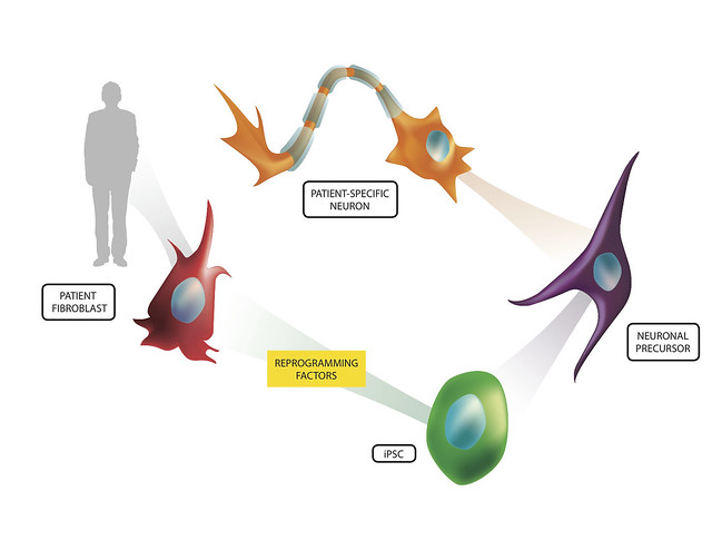
Stem cell research has generated promising and exciting results that could lead to treatment and cures for devastating diseases the medical community have been otherwise unable to treat. To some, the concept of stem cell treatment, though, was an ethical dilemma that shut the door on the seemingly endless possibilities these pluripotent cells could yield. Because of this, the discovery of induced pluripotent stem cells (iPSCs) was a very welcome discovery for the scientific community and the public. iPSCs are cells that once were well-differentiated (having a defined function in the body - i.e. skin cells), that are able to be coerced to 'going back in time' to obtain that pluripotent state seen in human embryonic stem cells (hESCs).

These iPSCs, once discovered, opened up those doors that ethical and moral questions shut for researchers. But, it was clear the iPSCs are different than hESCs and researchers had a fair bit of work in front of them to make sure that the iPSCs were at least comparable in the flexibility and thus utility of hESCs. This largely entailed experimenting with different environments and factors that the researchers would submit the cells to in order to create iPSCs. Eventually, researchers were able to derive iPSCs from more than just human skin cells (Yu et al, 2007), but other cells such as human urine cells (Zhou et al, 2012). As researchers have improved on these techniques, researchers like Morizane et al (2013) set out to investigate the quality of these iPSCs by injecting them into animals to investigate the utility of iPSCs as well as evaluate the benefits stem cells from one's own body may have on graft rejection or lack thereof.

In the study by Morizane et al (2013), the researchers used a primate model in order to evaluate the ability for autologous iPSCs and allogenic iPSCs to generate an immune response and engraft dopamine neurons in the primate midbrain. Like any other transplantation the immune system is an important player in the ability for the limb, cell, organ, etc. to engraft and become functional in the body. If recognized by certain immune cells in the body, the body can start attacking the transplant, reject it, and potentially damage the surrounding area in the process.
http://www.flickr.com/photos/lalande/6775578122/

Myeloid Dendritic Cells Enhance HIV Latency in T Cells


Diagram of the HIV virus

The Human Immunodeficiency Virus commonly known as HIV in the last 30 years has become a worldwide epidemic affecting approximately 23 million people with an additional 4 million cases annually.  This virus systematically disrupts and destroys the host immune system and is the direct precursor to Acquired Immunodeficiency Syndrome or AIDS.  Despite years of intense scientific research there is no cure or vaccine for HIV.  The current form of treatment is continual use of anti-retroviral drugs which can help to keep HIV at bay but are expensive and have side effects.  The virus itself invades a number of different immune cells and inserts its DNA into the DNA of the cell causing it to produce more of the HIV virus.  The virus itself is also toxic to subsets of these cells known as CD4+ T cells which die after the virus has replicated within the cell.  The destruction of these CD4+ T cells mediates the inability of the immune system to fight off other pathogens. 
                A major problem in combating HIV is that after the virus infects a cell it sometimes lays dormant in the cells to be replicated later.  In this case the virus is not immediately killing the cell but it in the meantime escapes normal processes of destruction.  This viral latency allows the virus to remain in the immune system undetected and escape destruction by other immune cells or anti-retroviral therapy.  Despite this common occurrence, the mechanisms underlying this process are relatively unknown.  However, a recent paper has partially uncovered how some CD4+ T cells are latently infected with the HIV virus.  A paper published this month by Evans et al. has demonstrated the myeloid dendritic cells are responsible for the latent infection of inactivated CD4+ T cells.  In this study, researchers co-cultured inactive CD4+ T cells with different types of infected dendritic cells to see whether they would cause a latent infection and if so which dendritic cells specifically.  The resting CD4+ cells were given a green fluorescent protein which gave off a green light when the cells were actively infected with the HIV-like virus.  The researchers then separated out the non-glowing cells which were not actively infected and further examined those.  When these CD4+ T cells were stimulated a small percentage of them turned green when they had not before, indicating that they contained the virus previously but it was not active and thus the virus was in a latent state.  This process allowed scientists to culture the CD4+ T cells with other cells or chemicals to see what factors caused the cells to have an active or latent infection.  Given this, it was soon discovered that infected myeloid dendritic cells caused a latent infection but not other types of dendritic cells.  Myeloid DCs are cells which migrate in the body and encounter pathogens.  These pathogens are then engulfed, chopped up by enzymes within the cell, and represented on the surface of the cell so that T cells may encounter the antigen and form an immune response against it.   Intriguingly it was previously thought that chemicals released by DCs or the environment called cytokines might be playing a role in the process, however, when the chemicals from the DC-T cell interaction were removed and cultured with new CD4+ T cells they did not develop a latent infection indicating that the myeloid DCs must have a physical connection to the T cells for this process to occur.  The connection between the two is an immunological synapse which involves numerous molecular adhesion molecules such as LFA-1 or ICAM.  The same study found that when they blocked these adhesion proteins there was a reduced effect of infection latency among CD4+ T cells but it did not eliminate latent infection completely so more than these two adhesion molecules must be involved.  These would be new potential targets for anti-retrovirals or other drugs.
Depiction of HIV spread from a DC to a T cell

Saturday, December 14, 2013

Can a virus stabilize Multiple Sclerosis?

Multiple Sclerosis (MS) is a neurodegenerative disease in which the neurons in the central nervous system (CNS; brain and spinal cord) cannot communicate effectively. The root of the disease is the degradation of the sheath of fat that surrounds some CNS neurons (myelin) - thus disrupting the effectiveness with which the neurons can survive and signal to each other. This destruction of the neurons is largely thought to be a result of the body attacking itself (an autoimmune disease), and not originating from an outside bacteria or virus; this hypothesis is largely the result of mice models that show that injecting components of this myelin sheath into a mouse will make the mouse exhibit MS-like symptoms.
Ref: http://health.howstuffworks.com/diseases-conditions/musculoskeletal/multiple-sclerosis1.htm

Although it seems as if this disease's cause is well-known and can be modeled well in animals, effective therapies to MS have proven hard to develop - one reason for this is that MS is different in almost every patient as to when it is symptomatic or not. This variation is so great that MS is characterized into many types of the disease such as relapse-remitting MS (RRMS) which the patient experiences many symptoms for months and then relapses into a largely symptom free condition, only to repeat in the future. Further, secondary progressing MS (SPMS) is a type of the disease - when symptomatic - declines at a devastating rate. There are more types, such as some that do not relapse and a continual decline is observed. It has been hypothesized that the Epstein-Barr virus (EBV; commonly known to cause mononucleosis) has some influence on this stability of disease - although results have not been very conclusive. This variability in the disease, as you can assume, confounds scientists researching therapies and potential cures to this devastating disease.

In hopes of elucidating why this variability is so great in this disease and if EPV has a role in disease stability, Annunziata et al in Italy investigated the role of EPV-positive B cells in disease severity. By extracting B cells from patients with MS, they examined the spectrum of antibodies that were produced by the MS patients of varying disease type. Initially, they identified 7 monoclonal antibodies (mAbs) that were found to bind a specific epitope (105-120) in one of the components of myelin that surround the neurons, MBP (myelin-basic protein). They chose this epitope of MBP because these mAbs were detected in the more 'stable' MS patients - and hoped to find something unique in these patients' body responses to the disease. Further testing included myelin-reactive T cells from MS to be evaluated in environments of the mAbs. Interestingly enough, only 3 mAbs showed dose-dependent inhibitory effects to the T cells - which are thought to contribute to the damaging environment in MS.

Friday, December 13, 2013

NKD2G and Allergic Inflammation


It seems to me that almost everybody nowadays has either a food allergy, seasonal allergy, or both.  Therefore, it becomes more important to understand the mechanism of allergies so that we can better treat them.  This paper examined the effect of the natural killer (NK) cell receptor NKD2G on pulmonary inflammation caused by allergic reactions. 

Allergies are a type of type 1 immune hypersensitivity (HS1).  There are two stages of HS1: sensitization and effector.  During the sensitization stage, the allergen is gathered by dendritic cells which move to the lymph node where they present the allergen to naïve Th cells.  These Th cells then provide T-cell help to B cells which release homing cytokines for other leukocytes to follow.  Other immune cells produce IL-4, IL-5 and IL-13 which facilitate isotype switching to IgE antibodies.  These antibodies then bind to receptors on the surface of basophils and mast cells.  The effector stage is the second exposure to the allergen.  The IgE antibodies bound to mast cells recognize the allergen and cause degranulation of the mast cells which have different effects depending on the tissue in which this is occurring.1

This video might help you understand immune hypersensitivity better as well:

The first experiment performed examined how NKG2D regulated the pulmonary inflammation caused by house dust mites (HDM).  HDM extract was given to mice that lacked the NKG2D receptor (klrkl-/-) and compared to those that had the receptor (klrkl+/+).  As a result, klrkl-/- mice showed a greatly reduced inflammatory response when compared to klrkl+/+ mice.  Additionally, there was less protein exudate in the airway when measured in the bronchoalveolar lavage (BAL) and significantly diminished recruitment of neutrophils, eosinophils and lymphocytes  in klrkl-/- mice.  Furthermore, of the CD4+ T cells that were recruited in klrkl-/- mice, much less IL-4 and IL-13 was secreted proportionally when compared to klrkl+/+mice.2 

Thursday, December 12, 2013

Transglutaminase-2 and Celiac Disease

Celiac disease is an autoimmune disease that has become increasingly prevalent in recent years.   It is an immune reaction that occurs with gluten, which is a protein found in wheat, barley, and malt.  This reaction causes inflammation, which damages the small intestine.  You may be thinking that wheat, malt, and barley are in everything!  So, what do people who suffer from celiac eat?  As someone who suffers from celiac, I can tell you the transition to a gluten free diet wasn’t easy, but luckily there are a lot of good, gluten free alternatives that have been developed.  Even though celiac can be helped with dietary accommodation, it is still important to try to understand what is causes the autoimmune response in order to potentially develop a treatment for celiac disease someday.  Unfortunately, it is not well understood how the autoreactive B cells become activated in celiac, but it is known that celiac disease is associated with autoantibodies produced that are specific for the transglutaminase-2 (TG2) enzyme.  In the paper Transglutaminase 2-Specific Autoantibodies in Celiac Disease Target Clustered, N-Terminal Epitopes Not Displayed on the Surface of Cells, the authors Iversen et al. investigated the mechanism that controls the formation of the TG2 autoantibodies.


Transglutaminase-2 is an enzyme that is involved in the deamination of glutamine residues, during them into glutamic acid.  This process increases their affinity for the HLA molecules associated with the disease, thus increasing the reactivity of gluten.  The anti-TG2 autoantibodies are an important marker of celiac disease, but it is unknown how these autoantibodies are contributing the disease.  In the lab’s previous research, they have identified various monoclonal (target 1 particular antigen) TG2-reactive antibodies (mAbs), and they have found that the antibodies target distinct (but close or overlapping) regions.  They define these regions as epitopes 1-4 throughout the paper. 
           
In order to determine the specificity of the mAbs, they stained the small intestinal tissue sections with immunofluorescent dye in either wild-type or mice deficient in TG2.  There was only fluorescence (indicating mAb binding) in the tissues containing TG2, which suggests that the mAbs are highly specific to TG2.  They also attempted to determine whether the mAbs would react with other members of the transglutaminase family, TG3 and TG6, which have also been associated with celiac disease.  They did not find any reactivity, which indicated that autoantibodies against other transglutaminases in celiac disease are created in dependently than the anti-TG2 autoantibodies.

TG2 has both an open and closed conformation.  It was thought that the epitopes on TG2 targeted by the autoantibodies were conformational, it would be expected that the mAbs would differ in binding strength to the two conformations.  Using an ELISA assay, the authors used the natural conformational regulators of TG2, Ca2+ (open conformation) and GTP (closed conformation) to measure the binding strength of the mAbs in each of the conformations.  The binding affinity of the mAbs was increased by Ca2+, but decreased by GTP.  This suggests that in celiac disease the open conformation of TG2 is the one that is targeted by the autoantibodies.

In a recent study done by Simon-Vecsei et al. , they identified an epitope targeted by celiac disease serum autoantibodies.  The authors wanted to know if their mAbs would react with the newly discovered epitope.  They constructed a triple mutation in the epitope and measured the binding strength with the mAbs by ELISA.  These mutations resulted in loss in reactivity for the mAbs in the epitope 2 group and the epitope 3 group.  This suggests that while this epitope is one of the major targets of autoantibodies in celiac disease, it is not the only one since the mAbs in the epitope 1 and 4 groups do not interact with it. 

Tuesday, December 10, 2013

Pediatric Brain Tumors Exhibit Distinct Phenotypes: Should Therapies Be Individualized?

Tumors are never something to joke around about; they are dangerous, unpredictable, and they can even be fatal. They can appear nearly anywhere in the human body, at basically any point in time. In fact, one of the most dangerous habitats for a tumor is the brain. A complicated and mysterious part of the human body already, the brain controls almost everything, and a tumor appearing in this region can be devastating. They are difficult to remove, and they can have disastrous mental and physical consequences. Even worse, they can present themselves at any age. This makes children susceptible, and what could be worse than that? With around 1,500 children diagnosed a year, pediatric brain tumors are not the most common cause of death in children (1). However, they are the most common type of pediatric tumor, and they have the highest mortality rate over all other childhood cancers. Despite its reputation, this deadly disease has had no improvements regarding treatment methods in the past. Until recently even, the standard treatment was radiation exposure and chemotherapy, which were often coupled with horrible, debilitating side effects. Now, a new form of treatment has been devised due to its tumor specificity – immunotherapy.

"Types of Brain and Spinal Cord Tumors in Children." Johns Hopkins Medicine.
Johns Hopkins University, Hospital, and Health System, 2013. Web. 10 Dec. 2013.
Studies have shown that there have been positive correlations between host immunity and survival rate in children diagnosed with brain tumors. Still, immunotherapy has demonstrated to be largely ineffective due to the immunosuppression induced by brain tumors because it tampers with the immune system-supressing qualities of the exogenous therapy. Several scientists researched pediatric brain tumor types further, however, in order to better understand immunophenotypes – the immunological characteristics – of these tumors. They hoped to shed some light on the subject in order to be able to better treat these afflicted children and give them more of a fighting chance.
               
In Andrea Griesinger and her colleagues’ study, they measured the phenotype and frequency of tumor-infiltrating leukocytes in the four most common types of child brain tumors*(4). They began by surgically taking tumor samples from forty-two patients at the Children’s Hospital in Colorado; they also took five non-tumor samples for a control group. Then, the tissue samples were disaggregated and frozen, before they were eventually suspended, stained, and analyzed via a FACS analysis and a gene expression analysis (2, 3). The FACS analysis then sorted the variety of cells into two or more containers based on their fluorescence while the gene expression analysis quantified the expression levels of certain genes.
             

Can Antibiotic Exposure Influence Development of Allergic Diseases?


A recent study provides certain implications for an association between exposure to antibiotics at a young age and the development of allergic diseases, primarily asthma, in early childhood.1


            At some point, most people have gone to the doctor’s office and left with a prescription for an antibiotic. In today’s world, antibiotics have developed a connotation as a medicine that can ward off all sorts of sicknesses, which is only partially true. What many people don’t realize is that antibiotics are strictly useful for fighting bacterial infections and will have no effect on viral illnesses.* Although antibiotics have saved countless lives since the discovery of penicillin, there are some concerns about their use.

There two main negative consequences of using antibiotics more liberally than in the past: some unhealthy bacteria have increased resistance to treatment and administration of antibiotics can lead to decreased levels of healthy bacteria. The first consequence relates to overprescribing antibiotics for patients that may not be suffering from a bacterial infection. Every time a person takes antibiotics, he or she increases the likelihood that bacteria in the body will become resistant, which makes it difficult to treat later infections.#

Commensal Bacteria
The second consequence, which directly relates to the study in question, has to do with the healthy bacteria that reside in the human gastrointestinal tract. When antibiotics are introduced to the body during infancy, a critical period for the development of the immune system, disruption to gut microflora can occur.1 This could possibly predispose patients to the development of an allergic phenotype. Research shows that disruptions in the normal growth of gastrointestinal bacteria can prevent regulatory T cells from properly dampening the immune system’s response to respiratory allergens.$  For more information, click here. Reduced diversity of microbes in infant excrement has also been connected to an increased risk of allergic diseases late in childhood.2

Allergic diseases develop when a person's immune system becomes sensitized to a normally harmless antigen. Type I hypersensitivity is a category of allergic reaction in which CD4+ Th2 cells that interact with these antigens stimulate B-cells to produce Immunoglobulin E (IgE) antibodies. These antibodies will then mark the specific antigen for destruction by other immune cells.+ Once the individual has been initially exposed and developed the specific antibodies, a subsequent exposure to the allergen will result in an allergic reaction. For more information, click here.

Mechanisms of Allergic Response

Monday, December 9, 2013

Autoimmunity, Rheumatoid Arthritis, and Abatacept


                        Figure 1: Example of rheumatoid arthritis effects.

You’ve likely heard of rheumatoid arthritis (RA), an autoimmune disease that results in a chronic, systemic inflammatory disorder. More than 2 million adults in the United States suffer from this disease, with women being two to three times more likely to develop it than men (UCSF Medical Center 2013; http://www.ucsfhealth.org/conditions/rheumatoid_arthritis/). The disease can occur at any age, although it typically affects those over 40 years old (Mayo Clinic 2013; http://www.mayoclinic.com/health/rheumatoid-arthritis/DS00020). RA is caused by an autoimmune attack on antigens expressed in the synovial tissue and cartilage of joints. Wrists, fingers, knees, feet, and ankles are most commonly affected. Early phase symptoms are characterized by morning stiffness in the affected joints. Over time, inflammation, cartilage destruction, and bone erosion may lead to deformations and crippling. The general explanation for these effects is as follows. The immune system attacks the synovium, which is the lining of the membrane that surrounds joints. Inflammation ensues which results in the thickening of the synovium and can eventually lead to the destruction of the cartilage and bone within the joint. The tendons and ligaments that hold the joint together also weaken and stretch, and can suffer degradation as a result of proteases (enzymes that break down proteins) secreted by activated macrophages (phagocyte meaning that they engulf solid particles). In this fashion, the joint gradually loses its shape and alignment, leading to deformities and crippling.


                            Figure 2: RA commonly affected joints and impact on joints.

So, more specifically, how does this all happen? Activated macrophages and DCs extravasate (essentially escape from a blood vessel into tissues) into the joint and produce large amounts of pro-inflammatory cytokines (intercellular mediators), especially TNF. The blood vessels in the inflamed joint take on the characteristics of high endothelial venules (HEVs), which specializes them for lymphocyte (small white blood cell) extravasation (Villani 2012; http://www.ipbs.fr/?High-endothelial-venules-HEVs). The inflammation is perpetuated by the eventual infiltration of the joint by CD4+ Th effectors (white blood cells that assist other immune cells) and CD8+ CTLs (white blood cells responsible for causing cell death of infected/damaged cells), which produce cytokines like TNF and IL-17. RA synovial tissues also contain “ectopic” germinal centers (sites where mature B lymphocytes proliferate, differentiate, and mutate and switch the class of their antibodies), meaning that these structures have developed in the wrong tissue. Plasma cells in these abnormal germinal centers produce autoantibodies (an antibody formed in response to and reacting against an antigenic constituent of its own tissues) directed against antigens (substance that induces an immune antibody response) in the synovial membrane and cartilage. Common markers of RA include the presence of rheumatoid factor and anti-citrullinated protein antibodies (ACPA) in the serum. These markers represent autoantibodies that have significant diagnostic values (da Mota et al. 2012; http://www.ncbi.nlm.nih.gov/pubmed/22187055).

 Figure 3: General immune overview of RA joint. Retrieved from Nutrition Remarks (2013) (http://www.nutritionremarks.com/2013/03/09/fish-oil-can-reduce-rheumatoid-arthritis-flu/)

ACPAs are autoantibodies present in the majority of patients with RA. They have proven to be useful biomarkers and allow for the diagnoses of RA at an early stage (da Mota et al. 2012; http://www.ncbi.nlm.nih.gov/pubmed/22187055). During inflammation, in a process known as citrullination, arginine residues in proteins can be converted to citrulline ones (Suurmond et al. 2011; http://www.ncbi.nlm.nih.gov/pubmed/21339220). If this change significantly alters the shape of the proteins, they may be seen as antigens and an immune response will be generated. Autoantibodies are generated against these citrullinated proteins (including fibrinogen or vimentin for example), forming the basis of an autoimmune disease. It is important to note that rheumatoid arthritis patients can be either ACPA-positive or ACPA-negative, and this status can have a significant influence on the intensity and therapy of RA. In fact, ACPA-positive and ACPA-negative RA have been recognized as distinct disease sub-entities, which demonstrate significant differences with regards to HLA-association, genetic and environmental risk factors, disease phenotype, and treatment response. 

Thursday, December 5, 2013

See See How CCL19 Effects Eosinophilic Pneumonia


Eosinophils are granulocytes that contain basic granules (i.e. secretory vesicles) that kill large parasites and are linked to various forms of allergies.  Eosinophilic pneumonia (EP) is a broadly defined disease that is characterized by an infiltration of eosinophils in lung alveolar tissue.  EP includes Churg-Strauss Syndrome (a rare autoimmune disease), chronic EP and acute EP (the difference between the two is the presence of eosinophils in the blood/tissues and only the tissue, respectfully).  Individuals with EP are often found to have an increased concentration of macrophages and dendritic cells, important innate immune response mediators.  Symptoms include shortness of breath, weight loss, fever, and even respiratory failure, while causes range from parasitic infection, immune system dysfunction, medication, and environmental stimuli like tobacco smoke and dust.  Although symptoms can be serious, few is known about the cellular mechanisms behind EP, specifically macrophage and DC recruitment into the lungs.  Therefore in response, Nureki et al. investigated EP further by seeing if either, both, or neither CCL19 and CCL21 (molecules that attract motile cells with a specific receptor to a specific location) bound to CCR7 expressed on DCs and macrophages, homing them into the lungs.
In order to extract cells present in alveoli of patients with EP and control individuals as noninvasively as possible, the researchers performed Bronchoalveolar lavage (BAL).  BAL is a procedure in which fluid (BALF) in released into the lungs and recollected (via bronchoscope.  Once BALF was collected, cytokines/chemokines were measured by enzyme-linked immunosorbent assay (ELISA), which is used to measure the concentration of antigens via antibody (complementary binding molecules) detection.  Finally, levels of proteins on cell surfaces were detected by immunocytochemistry, a technique that uses antibody binding and further bound-antibody detection.

Implications of Chronic Alcoholism for HIV Infection

http://9wtf.com/uploads/Meme-Drink-Alcohol-res.jpg
Alcohol dependence is the most common form of drug abuse in the US, with around 7% of the population meeting criteria for alcoholism (Grant 1994). We all know that the effects of alcohol are wide-ranging, impacting both behavior and physiology; it impairs judgement, motor skill, etc. It has been known to weaken the immune system, and research in the past few years has linked the effects of alcohol to HIV infection. Common sense tells us that alcohol promotes risky behaviors, including those that increase the possibility of HIV infection (sex, drugs, etc.). Recent interest in binge drinking and HIV has produced data showing alcohol in a binge pattern changed the proportion of immune cells after SIV infection in rhesus macaques, a common animal model for HIV, and may even increase the disease course (Molina et al 2006, Poonia et al 2006). But researchers at the Scripps Research Institute have also showed that chronic alcoholism may generate microenvironments in the body that are more vulnerable to HIV infection.

http://img.gawkerassets.com/img/18r4y1modssl5jpg/k-medium.jpg
To accomplish this study, the researchers developed a protocol in which rhesus macaques self-administered alcohol orally, twice a day for 30 days before infection and during infection. To do this, they mixed alcohol and an orange flavored drink in different ratios, and conditioned the animals to drink the solution (replacing water with the alcohol solution for a short duration). The final concentration given to the animals was 6% alcohol, similar to the alcohol content of most beers, and animals drank enough alcohol per day to cause blood alcohol levels that in humans are greater than the legal driving limit and would decrease motor skill. When the animals were conditioned, they were infected with SIVmac251, a strain of SIV that induces high peak and steady viral loads and is also known to infect the CNS (Burdo et al 2005). SIVs, simian immunodeficiency viruses, are retroviruses that infect non-human primates and produces symptoms and changes in physiology that mirror those induced by HIV infection (it is generally believed that HIV originated from SIV crossing the species barrier to humans).