-->
Showing posts with label microglia. Show all posts

Wednesday, December 18, 2013

How your own skins cells will one day treat your Parkinson's Disease.

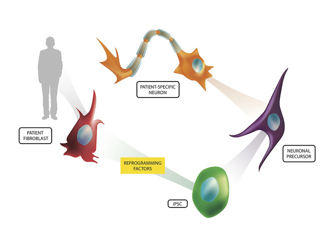
Stem cell research has generated promising and exciting results that could lead to treatment and cures for devastating diseases the medical community have been otherwise unable to treat. To some, the concept of stem cell treatment, though, was an ethical dilemma that shut the door on the seemingly endless possibilities these pluripotent cells could yield. Because of this, the discovery of induced pluripotent stem cells (iPSCs) was a very welcome discovery for the scientific community and the public. iPSCs are cells that once were well-differentiated (having a defined function in the body - i.e. skin cells), that are able to be coerced to 'going back in time' to obtain that pluripotent state seen in human embryonic stem cells (hESCs).

These iPSCs, once discovered, opened up those doors that ethical and moral questions shut for researchers. But, it was clear the iPSCs are different than hESCs and researchers had a fair bit of work in front of them to make sure that the iPSCs were at least comparable in the flexibility and thus utility of hESCs. This largely entailed experimenting with different environments and factors that the researchers would submit the cells to in order to create iPSCs. Eventually, researchers were able to derive iPSCs from more than just human skin cells (Yu et al, 2007), but other cells such as human urine cells (Zhou et al, 2012). As researchers have improved on these techniques, researchers like Morizane et al (2013) set out to investigate the quality of these iPSCs by injecting them into animals to investigate the utility of iPSCs as well as evaluate the benefits stem cells from one's own body may have on graft rejection or lack thereof.

In the study by Morizane et al (2013), the researchers used a primate model in order to evaluate the ability for autologous iPSCs and allogenic iPSCs to generate an immune response and engraft dopamine neurons in the primate midbrain. Like any other transplantation the immune system is an important player in the ability for the limb, cell, organ, etc. to engraft and become functional in the body. If recognized by certain immune cells in the body, the body can start attacking the transplant, reject it, and potentially damage the surrounding area in the process.
http://www.flickr.com/photos/lalande/6775578122/

Friday, December 16, 2011

A New Treatment Option for Alzheimer's Disease? IL Take It!

Alzheimer's disease (AD) is a devastating neurodegenerative disease that is, according to the National Institute on Aging, the most common form of dementia among older people. (1) Dementia is characterized by impairment in many mental faculties including, but not limited to: language, memory and perception. (2) Neuroinflammation is known, at this point in time, to play a role in AD. What exactly inflammation has to do with AD is currently a topic of much debate in scientific circles. An analysis of microarray data using microarray techniques identified 5 cytokines (immune system signals) to be important biomarkers of AD. (3) One of the cytokines identified in that study is IL-1, the production of which has been identified to be critical to the formation of the filamentous protein tangles that are a hallmark of AD. (4) This led a team of researchers at the University of California, Irvine to attempt inhibition of IL-1 signaling in a mouse model of AD with the application of an antibody directed against IL-1R (the receptor for IL-1) with the hope of improving AD symptoms. (5) This study is the focus of this blog post.
The researchers first did a behavioral study to test the efficacy of the antibody treatment on the cognitive abilities of their murine subjects. These mice are not your average laboratory mice; they have had three genes introduced into their cells that result in the accumulation of protein deposits and neuronal miscommunication that plague AD patients. (6) After treatment with the anit-IL-1R antibody, mice showed considerable improvement over non-treated mice in terms of their ability to navigate a water maze and to recall traumatic events (see figure below).

Neurodegeneration of Bacterial Meningitis

Streptococcus agalactiae (group B Streptococcus, GBS) and Streptococcus pneumoniae (SP) are the leading pathogens of bacterial meningitis in newborns and infants, respectively (1). GBS is a bacteria that lives in the gastrointestinal tract and can spread to the vaginal or rectal tissues in about 40% of women, where it can then be passed to the newborn through the birth canal (1). About one-third of all cases of meningitis in preterm infants and newborns are caused by GBS, while approximately half of meningitis in older infants is attributable to SP, in particular, in developing counties (2). Several studies have shown that neuronal vulnerability is extremely high in the first week of life and thus vulnerable to infection (3). During this time, there are crucial developmental processes that form the basis of the brain network. Without this development, brain function can be severely limited. During meningitis, the bacteria kill cells and release their fragments causing irreparable harm. Although mortality of meningitis in the very youngest age groups has dropped significantly, especially in developed countries, long-term neurological deficits such as deafness, blindness, cerebral palsy, seizures, hydrocephalus or cognitive impairment has remained virtually unchanged in 25%–50% of survivors(1).
The early immune response activates microglia (immune cells in the brain) and causes the influx of leukocytes into the cerebrospinal fluid (CSF). Leukocytes are essential for the elimination of replicating bacteria but also release toxic factors which add to neuronal damage (4). In infants, SP meningitis causes bacterial toxins such as the pore-forming pneumolysin and H2O2 (cause destruction of cells) have been identified as major neurotoxins. SP meningitis that lack pneumolysin and H2O2 has been shown to cause less neuronal damage (5). However, the contribution of pneumolysin and H2O2 to damage in neonatal animals has yet to be examined.
In a study done by Dr. Anja Reib at the Institute of Cell Biology and Neurobiology in Germany, they established a meningitis model of neonatal rats to study the neurodegeneration induced by GBS or SP. They looked at the role of the pore-forming cytolysins (similar to the pneumolysin in the SP bacteria) in triggering inflammation and neuronal damage.