-->

Friday, December 13, 2019

Enteroviruses and Autoimmunity


Enteroviruses are common throughout the United States. There are a variety of specific viruses that are categorized under this species [1]. Infection typically affects young children and causes fever, runny nose, sneezing, cough, and body aches. Enteroviruses are not usually dangerous and can be fought off by the child’s own immune system [2]. Type 1 diabetes (T1D) and islet autoimmunity are common in the United States as well. T1D is a chronic disease in which the pancreas produces insufficient insulin. Insulin is used by the body to help sugar enter the cells to be used to make energy. The exact cause of T1D is not entirely known. It is an autoimmune disease meaning that the body’s immune system usually attacks itself for unknown reasons [3]. Islet autoimmunity is the early stage of autoimmunity that occurs prior to a diagnosis of T1D [4] . The Vehik et al. paper is studying whether enterovirus can play a role in causing these autoimmunities.
            Vehik et al. wanted to study whether increased amounts of enterovirus infection is correlated with increased T1D or islet autoimmunity. They studied this by collecting fecal matter from 282 children with islet autoimmunity and 112 with T1D, plus age matched controls. The controls were children of roughly the same age who did not have either of these diagnoses. The stool from the controls was used to see what normal levels of virus shed in the fecal matter are. Shedding in terms of viruses is when after a person is infected with a virus the virus is present in bodily secretions (ex. Saliva, blood, stool) where it can be used to infect other people [5]. Then, the virus particles that were excreted in the stool were sequenced to determine the genetic code of the virus. These viruses were then analyzed to determine any correlation between virus particles being shed and disease outcome. Figure 1 shows the different viruses that were identified in the children’s stool. 55.8% of the samples were positive for mammalian viruses with the most prevalent being human adenovirus, parechovirus, Enterovirus A, and Enterovirus B.  In Figure 1C they broke down the percentages of Enteroviruses that were found in the stool and determined that the most prevalent forms of enterovirus were Enterovirus A and coxsackievirus. Coxsackievirus is what causes Hand, Foot, and Mouth disease in children [6]. Figure 1d looks at the likelihood of developing islet autoimmunity based after exposure to certain common human viruses based on the amount of virus positive stool samples. This analysis also showed a positive correlation with Human mastadenovirus F (HAdV-F). WHen HAdV-F was found in the stool, it correlated with lower levels of islet autoimmunity and T1D
Figure 1. Shows association of virus with disease prevalence


            This paper looked at global incidences of enterovirus correlation with islet autoimmunity and found results to be somewhat similar worldwide. Next, Vehik et al. did a longitudinal analysis where they looked at the amount of virus that was shed after initial infection to study how long virus could still be identified in stool. They identified virus shedding as prolonged when the virus being shed had 98% or more similarity in the genetic code (RNA). The results show that the same virus was being shed for an average of 6 months in children with islet autoimmunity and 4 months in the matched controls. Therefore, children with prolonged shedding of the same virus had higher odds for developing islet autoimmunity. If the virus with prolonged shedding was coxsackievirus B, then the likelihood of developing islet autoimmunity is even greater. This is seen in Figure 2 which studies the risk of islet autoimmunity based on viral infection frequency. It shows that prolonged shedding increases risk. Meaning, the longer the virus is replicating in the patient and the longer the patient is still producing infectious virus, the higher the likelihood of that patient developing islet autoimmunity is.
 
Figure 2. Shows risk of disease based on viral frequency.
Ultimately, the authors did some genome sequencing to look at mutations in the receptor that the virus binds to on the cell. This showed that certain changes seem to have a higher correlation with islet autoimmunity.This combined with the other results of the paper prove that prolonged shedding of the same virus are associated with higher risk of islet autoimmunity. The type and duration of the viral infection are both key factors in determining a child’s risk. They found a positive correlation with HAdV-F which appears to have a protective factor to prevent islet autoimmunity. HAdV-F binds to the same receptor as coxsackievirus B, indicating a potential for receptor competition which lessons the ability of coxsackievirus B to bind and cause infection. This study is mainly a correlational analysis, and much research is still needed to figure out how these viral infections cause autoimmunity.

Works Cited:
[1] Linden, Lonneke, et al. “Replication and Inhibitors of Enteroviruses and Parechoviruses.” Viruses, vol. 7, no. 8, Aug. 2015, pp. 4529–62. DOI.org (Crossref), doi:10.3390/v7082832.
[2] Non-Polio Enterovirus | About EV-D68 | Enterovirus D68 | CDC. 3 Jan. 2019, https://www.cdc.gov/non-polio-enterovirus/about/ev-d68.html.
[3] “Type 1 Diabetes - Symptoms and Causes.” Mayo Clinic, https://www.mayoclinic.org/diseases-conditions/type-1-diabetes/symptoms-causes/syc-20353011. Accessed 13 Dec. 2019.
[4] “Type 1 Diabetes: Autoimmune Reaction Successfully Halted in Early Stage Islet Autoimmunity.” ScienceDaily, https://www.sciencedaily.com/releases/2018/01/180104120349.htm. Accessed 13 Dec. 2019.
[5] “Viral Shedding.” TheFreeDictionary.Com, https://medical-dictionary.thefreedictionary.com/viral+shedding. Accessed 13 Dec. 2019.
[6]

The Ability of Hantaan Virus to Interfere with Host Immune Responses via the Manipulation of Autophagy

Paper: Wang, K., Ma, H., Liu, H., Ye, W., Li, Z., Cheng, L., ... & Zhang, F. (2019). The Glycoprotein and Nucleocapsid Protein of Hantaviruses Manipulate Autophagy Flux to Restrain Host Innate Immune Responses. Cell reports, 27(7), 2075-2091. [1]

Hantavirus is an infective agent relevant to public health concerns worldwide. As human-to-human transmission is rare, infection is primarily passed from rodent reservoir hosts into humans through aerosolization of mouse or rat bodily secretions [2]. Ultimately, the impacts of this infection pose great epidemiological threats through the syndromes they are linked to induce. In the Americas, for instance, “New World” hantaviruses primarily cause hantavirus pulmonary syndrome (HPS), which in turn leads to severe respiratory stress and high mortality rates. (Click
here to view the CDC interactive map of hantavirus incidents within the United States). Additionally, in Europe and Asia, “Old World” hantaviruses primarily manifest as hemorrhagic fever with renal syndrome (HFRS) upon infection. The major causative agent of this syndrome is Hantaan virus (HTNV), which is able to disrupt the permeability of kidney endothelial cells, ultimately resulting in acute kidney failure and mortality rates as high as 15% [2]. Although there are established vaccines and antiviral drugs to combat HTNV-induced syndromes in China and Korea, further public health interventions are being developed for hantavirus. Specifically, many clinical trials are currently being conducted for vaccines against causative agents of HPS, as therapeutic measures against these hantaviruses have yet to be successfully achieved in the United States and greater western hemisphere [3]. 

Therefore, many researchers continue to look into the pathogenic mechanisms of hantavirus infection with the hopes of uncovering new steps and proteins present in the viral pathway to target clinically. In this particular study, researchers were investigating the ability of HTNV to interfere with autophagy flux pathways facilitated by the host’s antiviral response to infection (see diagram A below and click link to cartoon) [4]. Specifically, this study aimed to investigate how HTNV is able to manipulate the autophagy pathway and which viral proteins are involved. HTNV is an enveloped virus containing a segmented genome composed of a single minus-sense strand of RNA. Therefore, viral proteins of particular interest to this study were the HTNV surface glycoprotein (Gn), as well as the nucleoprotein functioning to coat its genome within the viral particle (NP). These structures are depicted in diagram B below. 

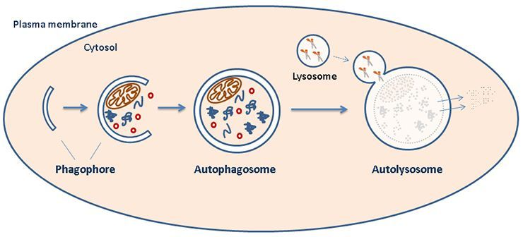
Diagram A: Host Cell Autophagy. At the start of the pathway, a double-membraned particle (phagophore) grows to surround specific cytosol contents to target for degradation. This structure matures into an autophagosome that ultimately fuses with a lysosome (forming an "autolysosome") to complete the degradation of contained substrates. [1]
Image source: https://www.med.uio.no/ncmm/english/news-and-events/profiles/images/cropped-illustration.jpg


Diagram B: Structure of Hantavirus. The virus contains three segments of (-)-sense ssRNA that each encode key viral proteins: small (S) encodes the nucleocapsid to coat the genome, medium (M) encodes the surface glycoproteins, and large (L) encodes the viral polymerase needed for replication. [1]
Image source: https://jasn.asnjournals.org/content/jnephrol/16/12/3669/F1.large.jpg

To first verify that HTNV infection acts to both induce and manipulate autophagy flux, researchers analyzed various known indicators of the pathway. Specifically, human umbilical vein endothelial cells (HUVECs) were infected with HTNV at various multiplicities of infection (amount of viral particles per cell) and immunoblots were performed 6 and 24 hours after infection. As shown below, two key indicators analyzed for autophagy were p62 and LC3B. The p62 protein is a receptor for substrates that are being sent for degradation, while LC3B is a microtubule-associated protein that acts within the selection process of this cargo [5; 6]. It is important to look at these two proteins together, as they have been found in previous studies to bind to one another and become degraded as a result of autophagy flux completion [7]. Therefore, the decrease of both p62 and LC3B at increased MOIs indicates that at 6 hours post infection (hpi), HTNV was indeed inducing complete autophagy flux. This is further supported by the increase across rising MOIs within Beclin1 levels, as this protein is an established indicator of autophagy initiation. However, at 24 hpi, these indicators did not demonstrate the same response to increased levels of HTNV infection, implying a disruption of host autophagy.

Figure 1B: HTNV induces host autophagy at early stages of infection. Levels of proteins p62, LC3B, and Beclin1 at differential MOIs of HTNV were detected in HUVECs at 6hpi. Reduced p62 and LC3B, as well as increased Beclin1 at high MOIs indicate complete autophagy at this time point post infection.

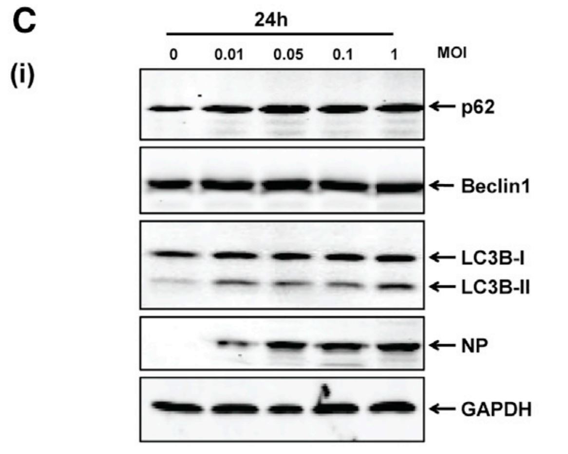
Figure 1C: HTNV blocks host autophagy at late stages of infection. Levels of proteins p62, LC3B, and Beclin1 at differential MOIs of HTNV were detected via immunoblot in HUVECs at 24hpi. Lack of changes in expression at high MOIs indicate incomplete autophagy at this time point post infection.

After visualizing these cells via electron microscopy, these findings were further supported by quantifying the number of autophagosomes as compared to autolysosomes at both time points. At 6 hpi, cell cultures exhibited more autolysosomes (the last step of autophagy flux in which substrates are degraded). Whereas at 24 hpi, cultures showed an accumulation of autophagosomes (an intermediate step demonstrating incomplete fusion with lysosomes). This prompted researchers to conclude that despite HTNV’s induction of autophagy at early stages, the virus is somehow able to manipulate the pathway to be incomplete at later stages of its infection.

In hopes of uncovering the viral mechanism beneath this manipulation, HEK293T cells were transfected with various proteins of HTNV and levels were detected via immunoblot. Of particular interest to researchers was the resulting levels of the viral surface glycoprotein (Gn) and nucleoprotein (NP). As shown below, with Gn expression alone, decreased p62 and LC3B levels indicated complete autophagy, just as they had in the prior experiment. An additional immunoblot also demonstrated that with decreased levels of Gn, there was a loss of this p62/LC3B response, indicating that Gn may be able to drive autophagy flux upon infection. However, with the addition of NP expression to that of Gn, there was a noticeable loss in the robust nature of this Gn-induced autophagy. This suggested that NP was somehow interfering with Gn’s ability to complete autophagy a hypothesis supported further by subsequent detection of autophagy progress via fluorescence microscopy. Using a reporter construct that is able to induce a color change from yellow to red when autophagosome to lysosome fusion occurs, increased yellow fluorescence was detected in Gn-NP expressing cells. This supported the conclusion that NP was blocking Gn at the particular autophagy step of fusion.

Figure 3A: Gn drives autophagy flux, unless inhibited by NP. Levels of autophagy indicators (p62 and LC3B) were detected via immunoblot in HEK293T cells transfected with HTNV viral proteins. Reductions in LC3B and p62 with Gn expression alone suggest the occurrence of complete autophagy, which is less robust in cells expressing NP+Gn.   

To build upon their findings that HTNV infection and viral Gn expression are able to induce autophagy flux within the host, researchers then aimed to determine the type of autophagy occurring as well as the mechanism of action. Given previous findings of the impact of viral infection on mitochondria within host cells, they investigated the possibility of the observed autophagy being the preferential degradation of mitochondria, termed “mitophagy” [8]. This hypothesis was supported via electron microscopy and flow cytometry of HUVECs that were either infected with HTNV or transfected to express Gn. Specifically, electron microscopy allowed researchers to visualize mitochondrial swelling within these cells, while the ability of flow cytometry to measure mitochondrial membrane potentials further demonstrated disturbed mitochondrial function under these conditions. 
Drawing again upon previous findings, researchers hypothesized that this mitophagy may be facilitated via the interaction of Gn with LC3B, the protein from their first experiments that functions in autophagy substrate selection. After identifying a particular sequence within the C-terminus of Gn that commonly binds with LC3B in other protein-protein interactions, mutants of the HTNV Gn were created by the researchers. Two mutants lacking the LC3B-binding motif (YRTL) were unable to cause the same level of mitochondrial damage as compared to full-length Gn and truncated mutants still able to interact with LC3B, as shown in the figure below. In addition, further investigations into Gn protein interactions revealed that the viral protein may also facilitate mitophagy through binding to a translation elongation factor associated with mitochondria (TUFM). Specifically, immunofluorescence imaging showcased colocalization of the proteins, and immunoblot revealed that TUFM knockdown cell lines exhibited significantly attenuated expression of mitophagy indicators. From these findings, a conclusion was made that Gn interacts with both the autophagy-associated protein (LC3B) and the elongation factor (TUFM) to trigger the degradation of mitochondria in infected cells.

Figure 4H: Loss of mitochondrial damage with disrupted Gn-LC3B interaction. HUVECs transfected with HA-tagged Gn mutants were imaged using a Mitotracker dye and cells with fragmented mitochondria were counted. Mutants lacking an LC3B binding motif (GnΔ35 and GnΔ42) showed decreased mitochondrial damage. 
Given this potential role in mitochondrial disruption, the question was then asked if HTNV Gn is also capable of interfering with the Mitochondrial Antiviral-Signaling Protein (MAVS) and its effects further downstream on the host innate immune system. Upon transfection of HUVECs to overexpress Gn, an immunoblot showing decreased MAVS levels led the researchers to claim that the viral protein does act to disrupt this antiviral signaling protein. Interestingly, looking at this result within cell lines depleted of the TUFM protein actually showed a restoration of MAVS levels, despite overexpression of Gn.The researchers in turn concluded that TUFM is a factor required for the degradation of mitochondria by HTNV Gn. Next, to look at subsequent impacts of HTNV infection on innate immunity, researchers then tracked the activity of reporter genes being expressed via promoters of either type 1 interferon (IFN) or the IFN-stimulated response element (ISRE). Interferons are a key indicator for the innate immune response to infection, as they are proteins released upon viral invasion and activate many genes via interactions with sequences such as the ISRE. Bringing the Gn mutants from prior experiments back into use, those unable to bind to LC3B (due to deletions or mutations of their YRTL motif) showcased significantly decreased IFN and ISRE reporter activity. At this point in their study, researchers concluded that at early infection HTNV Gn is able to facilitate complete mitophagy via its interactions with LC3B and TUFM, a process that in turn disrupts the host’s innate immune response to produce interferon. 
However, it is important to note that an ultimate consequence of this complete autophagy is, ironically, the eventual degradation of HTNV Gn. Not only is this an issue for progeny viruses being produced within the host cell, which need this glycoprotein to go off and infect other cells, but also this limits the suppression of MAVS and interferon antiviral responses. Therefore, the need for Gn to be maintained brings us back to two of the researchers' earlier findings: that HTNV infection leads to incomplete autophagy at late stages of infection and that HTNV NP has a role in blocking Gn autophagy (refer back to Figures 1c and 3a above). After recognizing via immunofluorescence that NP colocalizes with LC3B at later stages of infection, researchers hypothesized that NP is disrupting autophagy flux via preventing LC3B binding to Gn. Co-immunoprecipitation assays that utilized antibodies against a tag on the NP protein to detect interactions with LC3B, verified that this physical interaction was indeed occurring. Furthermore, additional co-immunoprecipitation assays revealed another key interaction between NP and a SNARE protein, SNAP29. Like other SNARE proteins, SNAP29 typically interacts with a syntaxin (Stx17) to drive the fusion of two membranes, which in this case are those of the autophagosome and lysosome. However, as shown below, researchers argue that NP’s binding to SNAP29 resulted in a lessened interaction of the protein with Stx17, thus indicating incomplete fusion, and in turn, the incomplete autophagy seen at late stages of HTNV infection. 
Figure 6G: HTNV NP disrupts SNAP29-Stx17 interaction. HEK293T cells were transfected with tagged Gn and/or NP HTNV proteins. Levels of coimmunopreciptation between SNAP29 and Stx17 were reduced in cells expressing both Flag-Gn and Myc-NP, as compared to cells expressing Flag-Gn alone.

Graphical Conclusion of Findings
To conclude their study, Wang et al. hoped to apply these findings to in vitro and in vivo models of HTNV infection via the application of autophagy inhibitors. Their reasoning behind this experiment was to interfere with autophagy early in infection, thereby limiting Gn’s ability to drive HTNV replication within hosts. Interestingly, infected mice showed increased levels of interferon, decreased organ damage, and reduced viral loads as a result of this treatment. Despite the benefits of this potential HTNV treatment, however, it is hard to imagine an autophagy inhibitor without a plethora of side effects. For instance, one concern of autophagy inhibitor-based treatments in the past has been potential neurodegeneration [9]. Thus, next steps to determine the viability of this treatment would be to track the long-term impact of their application, especially since the antiviral effects of the inhibitors in this study were only seen for 4 days post infection. In addition, a number of the conclusions within this paper could use further support. For instance, in Figure 6G above, the conclusion that NP significantly prevents SNAP29 binding to Stx17 is questionable, as there is still a clear interaction detected in the presence of NP. Perhaps further experiments, such as immunofluorescence and pull-down assays, are necessary to further support this claim. Additionally, it is still unclear how the virus is able to differentiate between Gn-induced autophagy and NP-induced incomplete autophagy. As mentioned by the researchers, the fact that Gn (as a surface glycoprotein) is a first contact to the host cell could be facilitating this early autophagy flux, before NP has an impact. Thus, a future study could possibly look into how applying HTNV infection to cells already transiently expressing viral NP would impact the completion of autophagy at early stages of infection. With these future considerations in mind, it is exciting to see such comprehensive research being done on a virus still in need of effective treatments worldwide. 

References

[1] Wang, K., Ma, H., Liu, H., Ye, W., Li, Z., Cheng, L., ... & Zhang, F. (2019). The Glycoprotein and Nucleocapsid Protein of Hantaviruses Manipulate Autophagy Flux to Restrain Host Innate Immune Responses. Cell reports, 27(7), 2075-2091.
[2] “Hantavirus.” (2019). Centers for Disease Control and Prevention. Retrieved from  https://www.cdc.gov/hantavirus/index.html
[3] Brocato, R. L., & Hooper, J. W. (2019). Progress on the prevention and treatment of hantavirus disease. Viruses, 11(7), 610.
[4] Klionsky, D. J. (2018). Why do we need autophagy? A cartoon depiction. Taylor & Francis Group. 
[5] Lamark, T., Kirkin, V., Dikic, I., & Johansen, T. (2009). NBR1 and p62 as cargo receptors for selective autophagy of ubiquitinated targets. Cell cycle8(13), 1986-1990.
[6] Barth, S., Glick, D., & Macleod, K. F. (2010). Autophagy: assays and artifacts. The Journal of pathology221(2), 117-124.
[7] Bjørkøy, G., Lamark, T., Brech, A., Outzen, H., Perander, M., Overvatn, A., Stenmark, H., and Johansen, T. (2005). p62/SQSTM1 forms protein aggregates degraded by autophagy and has a protective effect on huntingtininduced cell death. J. Cell Biol. 171, 603–614.
[8] Ding, B., Zhang, G., Yang, X., Zhang, S., Chen, L., Yan, Q., Xu, M., Banerjee, A.K., and Chen, M. (2014). Phosphoprotein of human parainfluenza virus type 3 blocks autophagosome-lysosome fusion to increase virus production. Cell Host Microbe 15, 564–577.
[9] Towers, C. (2017). Autophagy as a Therapeutic Target: The Double-Edged Sword. Novus Biologicals. Retrieved from https://www.novusbio.com/antibody-news/antibodies/autophagy-as-a-therapeutic-target-the-double-edged-sword

Thursday, December 12, 2019

Understanding Astrocyte Apoptosis: New Developments in Venezuelan Equine Encephalitis Virus Research

In Response to Dahal, B., Lin, S. C., Carey, B. D., Jacobs, J. L., Dinman, J. D., van Hoek, M. L., ... & Kehn-Hall, K. (2020). EGR1 upregulation following Venezuelan equine encephalitis virus infection is regulated by ERK and PERK pathways contributing to cell death. Virology, 539, 121-128.

The Venezuelan Equine Encephalitis Virus (VEEV) is a rare, but debilitating virus that can cause severe neurological symptoms in both humans and horses. VEEV is a positive-sense single-stranded RNA Alphavirus from the Togaviridae virus family. There are fourteen different strains of VEEV of which four, IA/B, IC, ID, and IE, can infect humans (Aguliar et al. 2011). The virus is spread to humans primarily through mosquito bites or contact with infected horses (Aguliar et al. 2011). VEEV infections occur in two stages. During the first stage, the virus infects the periphery organs, in particular, glands such as the lymph nodes and the spleen. During this stage, patients typically experience symptoms such as fever, vomiting, and diarrhea. The second, and more serious, stage of the infection begins when the virus progresses to the central nervous system (CNS). Infection in the CNS causes encephalitis, inflammation of the brain. If the patient develops encephalitis, they may experience convulsions or feel disorientated. Sometimes encephalitis can lead to death which occurs in around 1% of people infected with VEEV (Zacks and Paessler, 2010). Twelve countries in the Americas have reported outbreaks of this debilitating virus with the first documented outbreak occurring in Columbia in the 1950s (Aguliar et al. 2011).

VEEV is of concern to public health officials because of its potential use as a biochemical weapon of terror. During the Cold War, both the Soviet Union and the United States weaponized the virus by developing aerosol forms to use as a biochemical agent against enemy combatants (Davis, 1999). When the virus is in aerosol form, VEEV causes a more serious infection because it can directly infect the CNS through the olfactory system (Vogel et al., 1996). The potential for this virus to be used as a weapon of terror has prompted scientists to study the viral mechanism of the VEEV to develop drugs that prevent the debilitating neuropathology from occurring once the disease progresses to the second stage of the infection.

To gain a better understanding of how to address and prevent VEEV infection, scientists have begun investigating the changes in the brain that occur once VEEV has infected neurons. Astrocytes are one type of brain cells heavily targeted in the second stage of VEEV infection (Peng et al., 2013). Astrocytes are glial cells found in the brain that have many functions ranging from promoting CNS homeostasis to reducing brain inflammation following infection (Guttenplan and Liddelow, 2018). After VEEV infection, there is an increase in astrocyte apoptosis, cell-initiated cell death (Aronson et al., 2000). Dr. Kehn-Hall’s laboratory group at George Mason University recently released a paper in Virology which seeks to uncover the mechanism of VEEV infection that results in high levels of astrocyte apoptosis. The lab group sought to build off their previous research which showed that VEEV infection caused an increase in the level of early growth response 1 (EGR1) mRNA and proteins in astrocytes in a cancer cell line (Baer et al., 2016). EGR1 is an immediate-early gene associated with cell growth, survival, and apoptosis (Pagel and Deindl, 2011). In their 2019 paper, these researchers sought to replicate their findings in primary human astrocytes, because the primary astrocyte culture would be more reflective of what occurs in a human brain during VEEV infection. Additionally, the researchers were interested in uncovering which cellular pathways are responsible for the upregulation of ERG1 expression and, consequently, greater levels of cellular apoptosis.

Researchers in the Kehn-Hall lab first determined whether or not ERG1 expression is elevated in primary human astrocyte cultures after VEEV infection. They tested their hypothesis by infecting primary human astrocyte cell cultures with the TC-83 strain of VEEV. TC-83 is a live attenuated strain of the VEEV virus. Live attenuated strains are forms of viruses that can infect cells, but which have been modified so they are less virulent than unmodified strains of the viruses (Lauring et al., 2010). These strains of viruses are safer for laboratory use. Following infection, the scientists ran real-time quantitative polymerase chain reactions (RT-qPCR) to measure the respective amounts of ERG1 mRNA transcript levels in the primary human astrocytes after VEEV infection. RT-qPCR measures increases in levels of mRNAs of interest compared levels of control mRNAs, which are held at reliable levels in the cell. For this experiment, ribosomal RNA was used as a control for comparing levels of ERG1 mRNA level. The Kehn-Hall laboratory found a significant upregulation of EGR1 gene expression nine hours post-infection as displayed by the increase in fold change of EGR1 mRNA transcript (Figure 1). Thus, their RT-PCR analysis of VEEV infected primary human astrocyte cultures demonstrated that VEEV infected astrocytes have increased expression of the EGR1 mRNA.

Figure 1. Quantification of EGR1 Gene Expression Following VEEV Infection in Primary Human Astrocytes. EGR1 RNA levels in primary human astrocyte cell cultures incubated with either VEEV or a control MOCK injection were quantified at 3, 6, 8, and 18 hours post-infection. Analysis was completed with RT-qPCR. Significant upregulation of ERG1 transcripts was found in VEEV infected cells 9 and 18 post-infection.

After demonstrating that EGR1 levels were elevated in astrocytes following VEEV infection, the research group wanted to assess which internal cellular pathways were responsible for the upregulation in EGR1 expression. The researchers examined four pathways previously implicated in EGR1 upregulation: the extracellular signal-regulated kinase (ERK), p38 mitogen-activated protein kinase (p38 MAPK), phosphoinositide 3-kinase (PI3K) and the protein kinase RNA-like endoplasmic reticulum kinase (PERK) pathway. The researchers infected cells with VEEV and small kinase inhibitor molecules previously shown to inhibit either the ERK, p38 MAPK, PI3K, or PERK pathway. As before, the researchers quantified the levels of EGR1 expression with RT-qPCR. Their results are displayed in figure 2. The researchers found that inhibition of the ERK and PERK pathways caused a significant decrease in EGR1 gene expression in primary astrocyte cultures infected with VEEV when compared to control cell cultures, those treated with dimethyl sulfoxide (DMSO). Inhibition of p38 MAPK did not yield any significant changes in EGR1 expression when compared to control cells. Interestingly enough, treatment with PI3K inhibitors significantly increased the expression of EGR1 gene transcripts in astrocytes treated with VEEV. The researchers posited no explanation for why PI3K inhibitors triggered a rise in EGR1 transcripts, but in the future, the researchers should investigate this finding to determine why their results were inconsistent with previous papers that found that inhibition of PI3K triggered a decrease in EGR1 gene expression (Pagel and Deindl, 2011).

Figure 2. The Impact of Pathway Inhibition on EGR1 Transcript Expression. Researchers utilized RT-qPCR to assess the difference in EGR1 transcript levels following inhibition of the ERK, p38 MAPK, PI3K, and PERK pathways. Expression of EGR1 in cells treated with inhibitors was compared to the expression of EGR1 in cells treated with DMSO, a nonreactive solution. VEEV infected cells expressions were compared to MOCK control cells expressions. Researchers found a significant decrease in ERG1 expression following inhibition of the ERK and PERK pathways.

After identifying which cellular pathways were responsible for the upregulation of EGR1 after VEEV infection, the researchers examined whether inhibition of the PERK or ERK pathway would reduce astrocyte apoptosis and, consequently, increase cell viability. To test this hypothesis, the researchers used small interference RNAs (siRNAs) to target the ERK and PERK pathways in primary astrocytes before infecting the cells with VEEV. siRNAs target mature RNA sequences in cells by binding to commentary RNA base-pair sequences found in the targeted RNA sequence. siRNAs are designed in laboratories to fuse with specific mRNA and thus can reliably and consistently inhibit the expression of target molecules. Following binding, the siRNA directs the cleavage of the targeted RNA, effectively removing the RNA sequence from the cell (Lopez-Sierra and Esteller, 2012). Researchers transfected cells with siRNAs that targeted the ERK pathway, the PERK pathway and a negative siRNA which was used as a control. Cell viability assays measured ATP production in cells as a proxy measurement for cell viability; increased ATP production was used to demonstrated increased cell viability. The researchers also performed caspase 3/7 assays to assess the levels of apoptosis occurring in the primary human astrocyte cultures. Caspase 3/7 is a molecule released in late-stage apoptosis; an upregulation of this molecule in cell cultures indicates that a greater level of apoptosis is occurring in the cell. (Carrasco et al., 2003). Through these two assays, the researchers found that inhibition of both the ERK and PERK pathways increased astrocyte cell viability and decreased caspase 3/7 activity following VEEV infection (Figure 3). These results suggest that these two cellular pathways are significantly involved in triggering astrocyte apoptosis following VEEV infection.

Figure 3. Cell Viability and Caspase 3/7 Assays in VEEV infected Cells following ERK and PERK Inhibition. Levels of astrocyte viability and apoptosis following VEEV infection were analyzed through cell viability and caspase 3/7 assays. In cell cultures where ERK and PERK pathways were inhibited, there was a significant increase in cell viability and a decrease in caspase 3/7 activity. These results suggest that ERK and PERK pathways induce apoptosis in human astrocytes following VEEV infection.

Based on their experimental data, the researchers in the Kehn-Hall laboratory developed a model for the upregulation in EGR1 expression and resulting in apoptosis increase in astrocytes following VEEV infection (Figure 4). The researchers proposed that when VEEV infects the cell, the ERK pathway is directly triggered and begins to stimulate the expression of ERG1. While ERK is directly upregulated by VEEV, the virus’s proteins accumulate in the host cell’s endoplasmic reticulum which the cell recognizes as foreign proteins. The presence of foreign proteins causes the cell to activate the unfolded protein response (UPR) which stimulates the PERK pathway. The PERK pathway interacts with the ERK pathway and also increases the amount of EGR1 expression. Increased EGR1 expression ultimately leads to an increase in apoptosis in astrocytes.

Figure 4. Proposed Model for Human Astrocyte Apoptosis Upregulation Following VEEV Infection. Researchers at the Kehn-Hall lab proposed that VEEV infection directly triggers the ERK pathway and indirectly triggers the PERK pathway through activation of the cell’s unfolded protein response (UPR). Both of these pathways stimulate greater expression of the EGR1 gene which triggers apoptosis in human astrocytes.

Looking towards the future, the Kehn-Hall lab is interested in understanding why ERG1 expression causes an increase in cellular apoptosis. There are no experimentally verified mechanisms that explain why an upregulation of EGR1 causes an increase in apoptosis. The experimenters have theorized that ERG1 stimulates other genes in the cell which directly cause apoptosis, but they need to perform more research to support their theory on ERG1’s role in apoptosis. Beyond uncovering the role of ERG1 in apoptosis, the researchers need to develop an improved way to test the influence of ERK on ERG1 transcript expression and caspase 3/7 activity. In their RT-qPCR analysis of ERG1 transcripts and caspase 3/7 assays following ERK inhibition, the reduction in ERG1 expression and caspase 3/7 activity in the VEEV infected cells was remarkably similar to the reduction in ERG1 expression and caspase activity in the control cells. Thus, it is imperative for the researchers to perform additional tests on the influence of ERK inhibition on ERG1 expression to verify that the pathway does stimulate the gene expression following VEEV infection and not just generally cause a greater expression of all genes in astrocytes. On a broader level, the VEEV community should investigate how the rates of the disease could potentially increase with the dawn of climate change. VEEV is spread through mosquito vectors, and currently, climate change has caused a shift global temperature that facilitates the breeding of mosquitos (Aguliar et al. 2011). A global rise in the mosquito population could potentially trigger a massive surge in mosquito-transmitted illnesses such as VEEV. Scientists must continue to perform research on diseases like VEEV to prepare the global community for an exponential increase in mosquito-spread diseases.

References
Aguilar, P. V., Estrada-Franco, J. G., Navarro-Lopez, R., Ferro, C., Haddow, A. D., & Weaver, S. C. (2011). Endemic Venezuelan equine encephalitis in the Americas: hidden under the dengue umbrella. Future virology, 6(6), 721-740.

Aronson, J. F., Grieder, F. B., Davis, N. L., Charles, P. C., Knott, T., Brown, K., & Johnston, R. E. (2000). A single-site mutant and revertants arising in vivo define early steps in the pathogenesis of Venezuelan equine encephalitis virus. Virology, 270(1), 111-123.

Baer, A., Lundberg, L., Swales, D., Waybright, N., Pinkham, C., Dinman, J. D., ... & Kehn-Hall, K. (2016). Venezuelan equine encephalitis virus induces apoptosis through the unfolded protein response activation of EGR1. Journal of virology, 90(7), 3558-3572.

Carrasco, R. A., Stamm, N. B., & Patel, B. K. (2003). One-step cellular caspase-3/7 assay. Biotechniques, 34(5), 1064-1067.
Davis, C. J. (1999). Nuclear blindness: An overview of the biological weapons programs of the former Soviet Union and Iraq. Emerging infectious diseases, 5(4), 509.

Guttenplan, K. A., & Liddelow, S. A. (2018). Astrocytes and microglia: Models and tools. Journal of Experimental Medicine, 216(1), 71-83.

Lauring, A. S., Jones, J. O., & Andino, R. (2010). Rationalizing the development of live attenuated virus vaccines. Nature biotechnology, 28(6), 573.

Lopez-Serra, P., & Esteller, M. (2012). DNA methylation-associated silencing of tumor-suppressor microRNAs in cancer. Oncogene, 31(13), 1609.

Pagel, J. I., & Deindl, E. (2011). Early growth response 1—a transcription factor in the crossfire of signal transduction cascades.

Peng, B. H., Borisevich, V., Popov, V. L., Zacks, M. A., Estes, D. M., Campbell, G. A., & Paessler, S. (2013). Production of IL-8, IL-17, IFN-gamma and IP-10 in human astrocytes correlates with alphavirus attenuation. Veterinary microbiology, 163(3-4), 223-234.

Vogel, P., Abplanalp, D., Kell, W., Ibrahim, M. S., Downs, M. B., Pratt, W. D., & Davis, K. J. (1996). Venezuelan equine encephalitis in BALB/c mice: kinetic analysis of central nervous system infection following aerosol or subcutaneous inoculation. Archives of pathology & laboratory medicine, 120(2), 164-172.

Zacks, M. A., & Paessler, S. (2010). Encephalitic alphaviruses. Veterinary microbiology, 140(3-4), 281-286.

Possibility of a Transmissible Rabies Vaccine


Based On: Bakker, KM., Rocke, TE., Osorio, JE., Abbott, RC., … Streicker, DG. (2019) Fluorescent biomarkers demonstrate prospects for spreadable vaccines to control disease transmission in wild bats. Nature Ecology and Evolution, 3, 1697-1704.

            Many diseases that afflict humans, especially viral diseases, are transmitted into human populations from bats. In a recent study in Peru, researchers captured small portions of vampire bat colonies and applied orotopical gels to their fur in order to determine the ability of certain substances to be passed between bats.1 An orotopical gel is a substance that can be placed on the fur of the bat and will be transferred into other bats and the systems of the bat itself through oral contact and grooming. This study investigated the rates of orotopical transfer in order to computationally model how a potential rabies vaccine would spread through bat colonies in order to decrease the prevalence of the virus in bat populations. Vaccines are substances that are given to animals and humans in order to elicit an immune response that protects the organism from being pathologically infected by a certain virus or bacteria. Vaccines are the most effective way in decreasing the transmission of disease and they help protect the lives of animals and humans alike. Bakker et al. found that the application of orotopical gels to captured bats from colonies can possibly increase the rate of rabies vaccination and reduce the overall rates of the virus.
            This study was conducted because vampire bats are a common reservoir host of rabies. Rabies is a small RNA virus that can infect a wide range of mammals, including bats, rodents, canines, and humans.2 The disease infects many different cell types, including neurons of the central nervous system, causing inflammation in the brain and spinal cord that can lead to paralysis and death. Many animals will exhibit altered aggressive behavior prior to death which increases transmission of the virus through saliva entering broken skin of other organisms due to bites and scratches.3 The study was conducted with the hope to decrease the overall transmission of rabies to livestock, domestic animals, and humans by reducing the prevalence of rabies in the bat reservoir, or the animal that the virus persists in without causing symptoms.
First, the researchers wanted to determine what percentage of the bat colony would be exposed to their orotopoical gel biomarker, rhodamine B (RB), when it was applied to a certain proportion of the colony. RB causes hair follicles to fluoresce after ingestion, so was an ideal biomarker to track which individuals were attaining the substance in measurable quantities. It was found that in two different colonies, there was over 84% and 92% ingestion of RB in bats after application or transfer of the substance. This data suggests that each bat that had RB applied to their fur transferred the gel to an average of 1.45-2.11 untreated individuals depending on the colony. This would be equivalent to a 2.6 fold increase in population-level coverage. To further evaluate mechanisms of transfer, the group applied differently colored ultraviolet powder to furs of young and adult male and female bats. This allowed them to track which demographics were interacting with each other and if there would be an advantage of applying orotopical vaccines to a particular subset of the colony. They found that adult male bats had the greatest amount of contact with both sexes and should thus be targets as the recipients of the orotopical vaccine.
Fig. 1
Figure 1: Number of individuals vaccinated in different colonies following initial application of vaccine to a smaller proportion demonstrates increased rates. (a) shows the vaccinated individuals in the LMA5 colony, (b) shows the vaccinated individuals in the LMA6 colony, and (c) shows the vaccinated individuals in the LMA12 colony which migrated during data collection and is therefore excluded from this review. 

          After the initial collection of data from the smaller colonies, the research team performed computational models of how the rabies vaccines would alter the population susceptibility to infection and the prevalence of rabies within it. They found that if 20% of the colony were to have the vaccine applied, this would lead to an approximately 40% overall vaccination rate in the colony. Depending on the actual R0 of rabies, or the number of cases that would result from a single infected individual, the outbreak size would decrease by 45-75%. The researchers modeled their results for multiple values of R0 because estimates for this basic reproduction number range from as little as 0.6-2.0. Interestingly, the outbreak reduction was not found to be linear to the percentage of the population that initially had vaccine applied. It was found that after 30% of the population was vaccinated, with every 5% increase in vaccinations, there was a less than 5% reduction of outbreak size. This data will be useful for future vaccinations so the greatest rabies reduction can occur at the lowest overall cost.
Fig. 4
Figure 2: Outbreak size and duration was decreased with increasing levels of vaccination in bat populations through transmissible vaccines. (a) demonstrates that with different percentages of applied vaccine, outbreak size will decrease to a certain extent depending on basic reproduction number, (b) demonstrates the percentage protected based upon vaccination rates, (c) demonstrates the percent reduction in outbreak size after vaccine application, and (d) demonstrates the decreasing additional reduction after 30% have vaccine initially applied. 
Finally, the research team was interested in determining whether or not the transmissible vaccine would be more effective at reducing the chance of a rabies outbreak in bat colonies as compared with already existing measures that reduce risk. Currently, rabies outbreak reduction occurs through the application of “vampiricide,” a topical poison that results in the death of infected and infectable bats via oral ingestion. Not only is vampiricide controversial for ethical reasons, but it can also affect local ecosystems as well as possibly lead to the transmission of infections from one colony to another as bats seek other suitable mates due to declining numbers within their own colony. It was found that in preventative (application to prevent rabies invasion into historically uninfected population) and proactive (application in an area with low levels of rabies, but not in the colony itself) scenarios, vaccination was greatly favored over culling in order to reduce outbreak size and duration. In order for culling to be favored, 60% or more of the population would have to have vampiricide directly applied, which is an impractical proportion of the colony to capture and treat. Even in reactionary cases (application 60 days after a single rabies infection), vaccination was favored if less than 20% of the population could be captured and treated, which is a realistically attainable proportion.
Fig. 5
Figure 3: Transmissible vaccination was preferred over vampiricide in preventative, proactive, and reactionary scenarios. (a-c) demonstrate that vaccination reduces outbreak size and duration in preventative scenarios, (d-f) demonstrate that vaccination reduces outbreak size and duration in proactive scenarios, and (g,h) demonstrate that vaccination is favored for low percentages, but not high, in reactionary scenario.
This study is extremely important for the future of infections that are present in bats as a reservoir host. While this study specifically analyzed the reduction of rabies outbreaks in vampire bats in Peru, the same principle of transmissible vaccines may be applicable to other infections as well. Bakker et al. showed that transmissible vaccines would be effective at reducing the risk of rabies outbreaks in vampire bats, thereby reducing the risk that infections would be transmitted to livestock, domestic animals, and humans. Transmissible vaccines would allow for a greater percentage of wild-life populations to be vaccinated than can physically be captured and treated by humans. Transmissible vaccination also offers an alternative to culling methods, which are not a sustainable long-term solution ethically, ecologically, or economically for rabies reduction. One barrier that must be overcome is the current cost of large scale production of rabies vaccines, one reason that biomarkers were used rather than the vaccine in this initial study. As vaccine development improves, this method of transmissible vaccination for rabies, and other viruses carried by bats, will become favored to reduce the possibility of outbreaks. Until that time, orotopical vaccination should be used in combination with vampiricide in order to reduce the likelihood of a rabies outbreak in bat colonies that could have detrimental effects on humans.
 _________________________________________________________________________________

Bakker, KM., Rocke, TE., Osorio, JE., Abbott, RC., … Streicker, DG. (2019) Fluorescent biomarkers demonstrate prospects for spreadable vaccines to control disease transmission in wild bats. Nature Ecology and Evolution, 3, 1697-1704. 

Fooks, AR., Cliquet, F., Finke, S., Freuling, C., Hemachudha, T., … Banyard, AC. (2017). Rabies. Nature Reviews, 3(17091), 1-19. 

World Health Organization. (2019). “Rabies.” Health Topics.