-->
Showing posts with label inflammation. Show all posts

Thursday, December 5, 2013

See See How CCL19 Effects Eosinophilic Pneumonia


Eosinophils are granulocytes that contain basic granules (i.e. secretory vesicles) that kill large parasites and are linked to various forms of allergies.  Eosinophilic pneumonia (EP) is a broadly defined disease that is characterized by an infiltration of eosinophils in lung alveolar tissue.  EP includes Churg-Strauss Syndrome (a rare autoimmune disease), chronic EP and acute EP (the difference between the two is the presence of eosinophils in the blood/tissues and only the tissue, respectfully).  Individuals with EP are often found to have an increased concentration of macrophages and dendritic cells, important innate immune response mediators.  Symptoms include shortness of breath, weight loss, fever, and even respiratory failure, while causes range from parasitic infection, immune system dysfunction, medication, and environmental stimuli like tobacco smoke and dust.  Although symptoms can be serious, few is known about the cellular mechanisms behind EP, specifically macrophage and DC recruitment into the lungs.  Therefore in response, Nureki et al. investigated EP further by seeing if either, both, or neither CCL19 and CCL21 (molecules that attract motile cells with a specific receptor to a specific location) bound to CCR7 expressed on DCs and macrophages, homing them into the lungs.
In order to extract cells present in alveoli of patients with EP and control individuals as noninvasively as possible, the researchers performed Bronchoalveolar lavage (BAL).  BAL is a procedure in which fluid (BALF) in released into the lungs and recollected (via bronchoscope.  Once BALF was collected, cytokines/chemokines were measured by enzyme-linked immunosorbent assay (ELISA), which is used to measure the concentration of antigens via antibody (complementary binding molecules) detection.  Finally, levels of proteins on cell surfaces were detected by immunocytochemistry, a technique that uses antibody binding and further bound-antibody detection.

Wednesday, November 6, 2013

Inflammatory Bowel Disease: More Fun Than It Sounds


Inflammatory bowel disease (IBD) is a defined as inflammation of the intestines.  It comes in two forms: 1) Crohn’s disease (CD) which causes lesions in the entire wall of the bowel and 2) ulcerative colitis (UC) which is characterized as inflammation in the mucosal layer of the colon (hope this did not gross anyone out too much…).   Surprisingly though, the symptoms associated with IBD is NOT caused by a particular pathogen, but by the attack of one’s own immune system.  Therefore, IBS is characterized as an autoimmune disease.  Microbial translocation, where microbial products enter systematic circulation due to loss of barrier integrity of the intestines, has also been observed in IBD, as well as other disorders.  Although great strides have been made to the understanding of IBD, the roles of specific immune cells types are still not completely clear.  In order to better understand IBD, Funderburg et al. (2013) studied if the accompanying inflammation is linked to T-cell activation and thus microbial translocation. 

Funderburg et al. found several important results.  First, blood plasma samples were taken in individuals with IBD and soluble inflammatory markers were measured. CRP (a protein found in the blood during inflammation) and IL-6 (pro-inflammatory cytokine) levels both increased in IBD patients, which is not surprising because IBD is characterized by inflammation. 

Inflamed Colonic Tissue


Wednesday, October 16, 2013

Lack of Endogenous IL-10 Enhances Production of Pro-inflammatory Cytokines and Leads to Brucella abortus Clearance in mice


As we all know cytokines help us fight off pathogens by molecular signaling with the use of IFNs and ILs. IL-10 regulates helps balance pathogen clearance and immune response. Brucella abortus is a chronic inflammatory disease which can be found in humans as well as animals. Previous studies have shown that IL-10 is a critical cytokine for inflammatory response in the host and prevents damage. Another study by Fernandes and Baldwin, showed that anti-IL-10 resulted in up to 10-fold fewer bacteria in the spleen with mice infected with the same strain of Brucella. IL-10 directly affects IL-12 by down regulating it and presents a feedback loop which ensures there is not excessive inflammation. In this current study by Corsetti et. al. published in September 2013 he set out to find out the results of IL-10 in bacterial clearance, inflammatory response, and aftereffects of being infected with Brucella abortus.

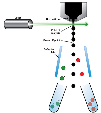
In order to test this, two sets of mice were studied, the wild type and the IL-10 KO (knock-out). Blood marrow cells were collected and cultured in DMEM in order to culture bacterial cells. After 10 days, the cells were infected with Brucella abortus and assayed for concentrations of IL-10, IL-12p40, or TNF-alpha. Brucella abortus strain 2308 was grown separately from the laboratory for 3 days and the mice were infected. Five animals from each group were examined at 1,2,3,6, and 14 weeks and spleens were removed. The spleens were plated and colonies were counted for cytokine analysis. To analyze the other factors such as IL-10 and IL-gamma, they stimulated the cells and unstimulated ones were used as negative controls. To test in vivo production of these factors, blood samples were taken and centrifuged, the supernatant was used for cytokine analysis. Real-Time RT-PCR was used on the splenocytes. FACS (Fluorescence activated Cell Sorter) is a type of flow cytometry in which cells in a heterogeneous mixture are sorted out.


A FACS analysis consists of many washes and separation method such as the one presented in the figure. The livers of the mice were also collected at each week period and stained with H and E. Granulomas (inflammation)  were measured using this method.

From this experimentation, we learned that wild-type infected with Brucella abortus presented increased production of IL-10 but none was found in the IL-10 KO mice as expected. IL-10 production was not only found in the dendritic cells but also in the splenocytes showing that it is produced in vivo and in vitro when infected. They determined that there was elevated proinflammatory cytokine production in IL-10 KO dendritic cells. Lack of IL-10 results in increased IL-12 and TNF-alpha production because it usually suppresses it. IL-10 KO cells resulted in less bacteria compared to the WT; by week 14 in the spleen there was no sign of Brucella abortus. All other factors were upregulated in IL-10 KO mice too. IL-10 KO mice has less granuloma in the liver as the infection time increased.

In conclusion, the knockout of IL-10 enhanced the the inflammatory response in Brucella abortus but could have adverse results in a different bacterial infection. The KO IL-10 led to reduction in liver pathology which is regulated by Treg cells and TGF-beta (increased in absence of IL-10). Further studies will be performed on these two key players.

I chose this article because I never thought of a cytokine having a better effect when being knocked out because I thought they all assisted in the immune response. Given this model was performed in mice, it’d be interesting if the same results would come from a human experiment. I believe it’s important to know these effects because it can help in cures of these disease and a key hallmark of this one is inflammation so this may work on other types of infection and even cancers.

Sources:


Thursday, October 3, 2013

Plasmacytoid dendritic cells: Friend or Foe?


Plasmacytoid dendritic cells (look a lot like plasma B cells but express different proteins on their cell surface) are important interferon-producers in the presence of viral RNA or DNA (an interferon is a cytokine, or a signaling protein released in response to a pathogens).  pDCs are important in linking the innate (nonspecific) and adaptive (specific) immune response through increasing the function of natural killer and T cells, respectfully.

Much is still unknown about pDC function, though.  As discussed in Li et al., there is a debate on whether pDCs inhibit HIV/SIV replication or instead promote chronic immune activations and disease.  In other words, what is the role of pDCs in lentiviral (long incubation period) infections?  Using SIV (Simian immunodeficiency viruses)-infected rhesus monkeys, Li et al. tried to determine the systematic distribution of pDCs using a cell counting/sorting mechanism called flow cytometry.  They also looked their cytokine expression and affect on T cell activation upon infection. 

Monday, January 9, 2012

Keep the hygiene, lose the inflammation: TGF-β and helminthic therapy

            Incidences of autoimmune and autoinflammatory diseases, such as type 1 diabetes, rheumatoid arthritis, and inflammatory bowel disease, are increasing in the developed world, and the annual costs of treatment amount to billions of dollars in the United States alone (1). There are numerous factors that can account for this rise in prevalence, including genetic differences and environmental factors. One factor that has been gaining support, both correlatively and experimentally, is the hygiene hypothesis: basically, since we have eliminated many of the prominent childhood diseases, children are “too clean,” and thus their immune systems, rather than focusing on pathogens, are stimulated to attack “self” targets instead, or target innocuous factors leading to allergy. In other words, infection with some types of pathogens, particularly parasitic worms such as roundworms, flatworms, and hookworms, can dampen the inflammatory responses that underlie many of these autoimmune/autoinflammatory conditions. We have largely eliminated these infections, which are transmitted through unclean drinking water and soil, in developed nations, thus accounting for some of the rise in autoinflammatory diseases.
            Some suffering patients have taken this idea to the extreme, and have deliberately infected themselves with parasites to try to alleviate their symptoms. Although this treatment is highly experimental and there is not a lot of clinical data demonstrating efficacy, anecdotal evidence suggests that some patients have observed marked reduction in symptoms following “helminthic therapy.” Obviously, there are many problems associated with introducing parasitic organisms into people, so if scientists can determine how helminthic infection tamps down inflammatory responses, it could potentially lead to novel anti-inflammatory treatments that don’t involve the parasites themselves.
            At the core of this issue are two major categories of immune responses, named “Th1” and “Th2” responses for the types of helper T cells that facilitate them. Th1 responses are inflammatory in nature, and involve the activation of macrophages and killer (CD8+) T cells. These responses are elicited by the secreted cytokine interferon-γ (IFN-γ), and typically target intracellular pathogens such as viruses. Th2 responses are mediated largely by B cells, which make antibodies, and are elicited by the cytokine interleukin-4 (IL-4). Th2 responses target extracellular pathogens, such as parasitic worms, and the rationale behind the hygiene hypothesis is that by eliminating many of these Th2 pathogens, the immune system “skews” towards Th1 responses. This, then, leads to increased inflammation and the associated autoinflammatory diseases. However, other responses caused by helminth infection, including the generation of regulatory T cells (TRegs) and the secretion of immunomodulatory cytokines such as IL-10 and transforming growth factor-β (TGF-β), might also mitigate autoinflammatory disease. A recent paper by Hübner et al attempted to distinguish which of these mechanisms was responsible for the protection from autoimmunity accorded by helminth infection. They found that generation of a Th2 response was not required, but that the production of TGF-β was largely responsible for protection.

Saturday, December 17, 2011

Prion Diseases: Are Researchers Finally Close to Uncovering a Cure?

Prion diseases are no laughing matter, although it might have seemed that way if you were a visitor on the island of Papua New Guinea in the 1950’s. It was here that members of the Fore tribe were suffering from what they had dubbed the “laughing sickness” due to the strange uncontrollable bursts of laughter that accompanied the debilitating shivering that struck it’s victims. The source of the disease, which came to be known as kuru, was found to be transmitted through ingestion of brain tissue during cannibalistic burial ceremonies. Ingestion of the diseased victims brain tissues exposed tribe members to a structurally altered form of the normally expressed cellular prion protein (PrP) which could react with healthy forms of the proteins in their brains, perpetuating further structural transformations.

There are a number of diseases spread in this manner, termed transmissible spongiform encephalopathies (TSE). All TSE's, which include the well-known mad cow disease (Creutzfeldt-Jakob disease), are inevitably fatal and lead to neurodegeneration, often mediated through pathologic neuroinflammation. The inflammation seems to be primarily due to the robust activation of microglial cells (Yang et al., 2008). Microglial cells are akin to the brain’s garbage trucks, constantly collecting cellular debris from the extracellular environment. This behavior coincidentally makes them especially fit for detecting the presence of extracellular pathogens as well.

Before a cure can be developed, researchers must first uncover the cellular mechanisms underlying the microglial-medated pathologic neuroinflammation observed in the victims of TSE. PrP106-126 is the region of the PrP protein that has been shown to mediate inflammatory and pathologic signaling following structural alteration. A recent study was able to use this peptide to identify many of the proteins and signaling molecules, thus potentially uncovering future targets for pharmaceutical therapies.

You Are What You Eat: A New Therapeutic Approach to IBD

Virulent fimbria expression on bacterial mats in the absence and presence of Phloretin (Lee et al., 2011)


Many inflammatory bowel diseases (IBD) such as Chron's (CD) or ulcerative colitis (UC) are considered autoimmune diseases caused by the body's immune system attacking the gastrointestinal tract. This is thought to trigger colitis, the medical term used to describe colonic and often intestinal inflammation. Specifically, a failure to regulate T cell responses are believed to trigger the inflammation of the intestinal or colonic mucosa in these disorders.

Researchers have begun to suspect intestinal microflora inhabiting the mucosae play a significant role in the development of IBD and colitis. Patients suffering from IBD have been shown to have higher concentrations of mucosal bacteria that increase progressively with symptom severity. The mucosae are thin membranes that are found throughout the body in areas where internal organs and tissues are exposed to external environments. They possess unique immune systems that are tightly regulated and hyporesponsive, or unresponsive to most antigens, due to the high antigen loads they come into contact with (Neurath et al., 2007). Because the mucosae are so sensitive to inflammatory damage, disruption of the hyporesponsive state can have devestating effects.

Phloretin is a type of flavonoid found mainly in apples and strawberries that has antioxidative and anticarcinogenic properties, as well as biological roles in estrogen hormonal activity and cardiovascular disease prevention. If the roles of phloretin weren't already diverse enough, researchers have also recently found flavonoids can inhibit the biofilm formation of pathogenic E. coli O157:H7. Bacterial biofilms are aggregates of bacteria that aggregate together and adhere to a surface. This might explain another recent study that found phloretin is capable of reducing the expression of many inflammatory proteins and receptors in human colon epithelial cells.

Recently, this relationship between phloretin and biofilm formation was further investigated by a team of researchers hoping interested in whether phloretin's inhibitory effect on biofilm formation could have a therapeutic effect on patients of IBD (Lee et al., 2011).

Friday, December 16, 2011

A New Treatment Option for Alzheimer's Disease? IL Take It!

Alzheimer's disease (AD) is a devastating neurodegenerative disease that is, according to the National Institute on Aging, the most common form of dementia among older people. (1) Dementia is characterized by impairment in many mental faculties including, but not limited to: language, memory and perception. (2) Neuroinflammation is known, at this point in time, to play a role in AD. What exactly inflammation has to do with AD is currently a topic of much debate in scientific circles. An analysis of microarray data using microarray techniques identified 5 cytokines (immune system signals) to be important biomarkers of AD. (3) One of the cytokines identified in that study is IL-1, the production of which has been identified to be critical to the formation of the filamentous protein tangles that are a hallmark of AD. (4) This led a team of researchers at the University of California, Irvine to attempt inhibition of IL-1 signaling in a mouse model of AD with the application of an antibody directed against IL-1R (the receptor for IL-1) with the hope of improving AD symptoms. (5) This study is the focus of this blog post.
The researchers first did a behavioral study to test the efficacy of the antibody treatment on the cognitive abilities of their murine subjects. These mice are not your average laboratory mice; they have had three genes introduced into their cells that result in the accumulation of protein deposits and neuronal miscommunication that plague AD patients. (6) After treatment with the anit-IL-1R antibody, mice showed considerable improvement over non-treated mice in terms of their ability to navigate a water maze and to recall traumatic events (see figure below).

Tuesday, December 6, 2011

Regulatory T Cells in Patients with Whipple's Disease

Classical Whipple’s Disease (CWD) is a rare, multisystemic infection of the duodenal mucosa. Macrophages infected with Tropheryma whipplei, a gram positive bacterium, first attack the intestinal mucosa and then disperse throughout the body to the intestinal epithelium, capillary and lymphatic endothelium, synovium, heart, lungs, liver, brains, eyes and skin. Symptomatic manifestations are most commonly reported in the intestines, and include weight loss, diarrhea, and abdominal pain (Schijf et.al 394). Additionally, T.whipplei infection can spread to the brain and heart and cause inflammation of the heart muscle. [1]
Schinnerling and colleagues argue that host immune deficiency plays a major role in the persistence and systematic spread of CWD. The authors suggest that healthy adults generally display an efficient humoral, or antibody, and cellular immunological response that effectively combats T.whipplei. However, immunological defects are believed to weaken the immune response and enable the pathogen to successfully infect the host [2]. The defects present in the peripheral blood and the duodenal mucosa of CWD patient include, but are not limited to, impairment of T cell proliferation, and diminished Th1 reactivity.
T cells are a type of white blood cell that helps recognize and eradicate invading pathogens that cause disease. Th1 cells are a brand of T cells that specialize in intracellular pathogen disposal. Specifically, Th1 cells secrete IL-2, IFN­-y, and LT, which are cytokines, or signaling chemicals, that initiate a robust inflammatory response. A Th1 response is generally avoided in mucosal tissues because inflammation is an aggressive immune response that can damage the delicate tissue of the mucosa. Consequently, a Th2 response is initiated in the mucosa to avoid cell damage and effectively combat extracellular pathogens. Th2 cells secrete cytokines, such as IL-4, IL-5, and IL-10, to suppress a Th1 response and initiate antibody production. Antibodies neutralize pathogens without damaging surrounding host cells [3].

Wednesday, November 30, 2011

Better Treatments for Rheumatoid Arthritis Not Too Far Off


Rheumatoid Arthritis (RA) is an inflammatory autoimmune disease that most often targets peripheral joints, frequently resulting in pain, swelling, and possible deterioration and destruction of cartilage and bone located at the joint. RA afflicts more than 1.3 million people in the United States alone, and the disease is most prevalent among women and the elderly. For over 60 years, RA has been widely treated non-selectively with glucocorticoids (GCs), such as methotrexate, a type of steroid hormone, because of their unrivaled, potent anti-inflammatory effects. Despite the successful anti-inflammatory effects that GCs have on the joints of afflicted patients (in fact, GCs are successful in just about every type of inflammatory ailment), it is a costly treatment, and there are numerous, severe side effects due to their non-selective nature, including immunodeficiency, high blood sugar, and increased skin fragility and bruising, among others. Consequently, there is currently a necessity for research into possible steroid therapies better targeted to healing RA with greater efficiency and fewer adverse side effects.
When RA is treated with GC steroid hormones, the hormone binds to a glucocorticoid receptor (GR) located in host cells (and expressed nearly universally in all vertebrate cells). Once being bound by a GC, the GR then translocates into the host cell nucleus where it can act as a transcription factor, subsequently altering gene expression and helping to dampen the host immune response and reduce inflammation at the site of RA. The GR may carry out this function via two different modes of action: it may dimerize, or split into two separate subunits, and then bind to the gene promoter of GC-regulated genes, or it may remain as a single unit and interact with other DNA-bound transcription factors, thereby altering gene expression to induce anti-inflammatory function.
While it is well understood that GCs induce potent anti-inflammatory effects, the specific cells targeted by GCs and their underlying mechanisms are poorly characterized. Just yesterday, a study was published in the Proceedings of the National Academy of Science by Baschant and colleagues that identifies a potential mechanism by which GCs actually carry out their anti-inflammatory function. The paper suggests that the GR in T cells, which are cells that contribute to the inflammatory state in joints, is critical for the suppression of inflammation by GCs, and that the dimerization of GRs is necessary for the GC’s anti-inflammatory effects. To achieve this, the authors utilized a mouse model in which they used an antigen-induced arthritis (AIA) to mimic the severe inflammation in joints that is characteristic of RA, and serves as a model to examine the mechanism by which GCs carry out their function.