-->

Wednesday, December 18, 2013

The Role of CD40–CD154 Interactions in Autoimmunity and the Benefit of Disrupting this Pathway


Autoimmune disease is associated with adaptive immune component dysfunction such as the B cells and T cells. The cells somehow pass through peripheral tolerance and cause a self reaction. Autoimmunity is determined by genetics, environmental, and hormonal effects that contribute to the disease. Many autoimmune diseases have been studied and examined but the biology behind them are not completely understood.

A major pathway is the CD40-CD40 ligand (CD154) because it is needed to activate many adaptive immune cells such as DC's, B cells, and T cells. This interaction has multiple functions and as costimulatory molecules, they are upregulated in several autoimmune diseases for example SLE (systemic lupus ertyhematosus). It was thought that disabling this interaction could be a new avenue of therapy for autoimmunity.


Caption: The image to the left shows the basic interactions between immune cells in the body when trying to get rid of an antigen. The same interactions occur in autoimmune diseases where the body is attacking self antigens. In this picture, we can see the CD40-CD40L (CD154) interaction described above between a dendritic cell presents antigens and a CD4+ T cell which aims to help the immune response by releasing cytokines.


CD40 provides a help signal to dendritic cells to maturation. The duration of the signal assists in determining the function of the dendritic cells which is displayed in this study. There are various other proteins that can trigger dendritic cells to become activated and thus spark an autoimmune response such as heat shock proteins. These can skew a response using cytokines, interleukins, and different factors. In a mouse model, diabetes was induced when CD40 expression on bone marrow was necessary for heat shock protein induced activation. 

This reaction of CD40-CD154 is not only found in dendritic cells but it also impacts T cells by priming them, polarizing T cells to a certain response.


Caption: The basic polarization of T cells is shown in the following image with different cytokines determining the Th cell developed. 

Th (T helper) cells only express CD154 after they have been stimulated by an antigen which allow them to have this interaction of CD40-CD154; unfortunately over expression of CD154 can lead to autoimmune diseases. This not only occurs in T cells but in B cells as well. The CD40-CD154 interaction enhances CD86 expression on B cells in autoimmunity specifically SLE disease which is a hypersensitive reaction. CD86 contributes to the presentation of self-antigens to T cells thus inducing a negative immune response by the body. 

How your own skins cells will one day treat your Parkinson's Disease.

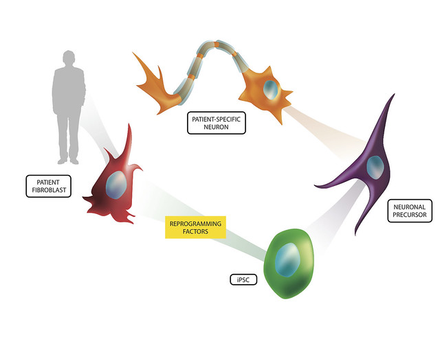
Stem cell research has generated promising and exciting results that could lead to treatment and cures for devastating diseases the medical community have been otherwise unable to treat. To some, the concept of stem cell treatment, though, was an ethical dilemma that shut the door on the seemingly endless possibilities these pluripotent cells could yield. Because of this, the discovery of induced pluripotent stem cells (iPSCs) was a very welcome discovery for the scientific community and the public. iPSCs are cells that once were well-differentiated (having a defined function in the body - i.e. skin cells), that are able to be coerced to 'going back in time' to obtain that pluripotent state seen in human embryonic stem cells (hESCs).

These iPSCs, once discovered, opened up those doors that ethical and moral questions shut for researchers. But, it was clear the iPSCs are different than hESCs and researchers had a fair bit of work in front of them to make sure that the iPSCs were at least comparable in the flexibility and thus utility of hESCs. This largely entailed experimenting with different environments and factors that the researchers would submit the cells to in order to create iPSCs. Eventually, researchers were able to derive iPSCs from more than just human skin cells (Yu et al, 2007), but other cells such as human urine cells (Zhou et al, 2012). As researchers have improved on these techniques, researchers like Morizane et al (2013) set out to investigate the quality of these iPSCs by injecting them into animals to investigate the utility of iPSCs as well as evaluate the benefits stem cells from one's own body may have on graft rejection or lack thereof.

In the study by Morizane et al (2013), the researchers used a primate model in order to evaluate the ability for autologous iPSCs and allogenic iPSCs to generate an immune response and engraft dopamine neurons in the primate midbrain. Like any other transplantation the immune system is an important player in the ability for the limb, cell, organ, etc. to engraft and become functional in the body. If recognized by certain immune cells in the body, the body can start attacking the transplant, reject it, and potentially damage the surrounding area in the process.
http://www.flickr.com/photos/lalande/6775578122/

Myeloid Dendritic Cells Enhance HIV Latency in T Cells


Diagram of the HIV virus

The Human Immunodeficiency Virus commonly known as HIV in the last 30 years has become a worldwide epidemic affecting approximately 23 million people with an additional 4 million cases annually.  This virus systematically disrupts and destroys the host immune system and is the direct precursor to Acquired Immunodeficiency Syndrome or AIDS.  Despite years of intense scientific research there is no cure or vaccine for HIV.  The current form of treatment is continual use of anti-retroviral drugs which can help to keep HIV at bay but are expensive and have side effects.  The virus itself invades a number of different immune cells and inserts its DNA into the DNA of the cell causing it to produce more of the HIV virus.  The virus itself is also toxic to subsets of these cells known as CD4+ T cells which die after the virus has replicated within the cell.  The destruction of these CD4+ T cells mediates the inability of the immune system to fight off other pathogens. 
                A major problem in combating HIV is that after the virus infects a cell it sometimes lays dormant in the cells to be replicated later.  In this case the virus is not immediately killing the cell but it in the meantime escapes normal processes of destruction.  This viral latency allows the virus to remain in the immune system undetected and escape destruction by other immune cells or anti-retroviral therapy.  Despite this common occurrence, the mechanisms underlying this process are relatively unknown.  However, a recent paper has partially uncovered how some CD4+ T cells are latently infected with the HIV virus.  A paper published this month by Evans et al. has demonstrated the myeloid dendritic cells are responsible for the latent infection of inactivated CD4+ T cells.  In this study, researchers co-cultured inactive CD4+ T cells with different types of infected dendritic cells to see whether they would cause a latent infection and if so which dendritic cells specifically.  The resting CD4+ cells were given a green fluorescent protein which gave off a green light when the cells were actively infected with the HIV-like virus.  The researchers then separated out the non-glowing cells which were not actively infected and further examined those.  When these CD4+ T cells were stimulated a small percentage of them turned green when they had not before, indicating that they contained the virus previously but it was not active and thus the virus was in a latent state.  This process allowed scientists to culture the CD4+ T cells with other cells or chemicals to see what factors caused the cells to have an active or latent infection.  Given this, it was soon discovered that infected myeloid dendritic cells caused a latent infection but not other types of dendritic cells.  Myeloid DCs are cells which migrate in the body and encounter pathogens.  These pathogens are then engulfed, chopped up by enzymes within the cell, and represented on the surface of the cell so that T cells may encounter the antigen and form an immune response against it.   Intriguingly it was previously thought that chemicals released by DCs or the environment called cytokines might be playing a role in the process, however, when the chemicals from the DC-T cell interaction were removed and cultured with new CD4+ T cells they did not develop a latent infection indicating that the myeloid DCs must have a physical connection to the T cells for this process to occur.  The connection between the two is an immunological synapse which involves numerous molecular adhesion molecules such as LFA-1 or ICAM.  The same study found that when they blocked these adhesion proteins there was a reduced effect of infection latency among CD4+ T cells but it did not eliminate latent infection completely so more than these two adhesion molecules must be involved.  These would be new potential targets for anti-retrovirals or other drugs.
Depiction of HIV spread from a DC to a T cell

Saturday, December 14, 2013

Can a virus stabilize Multiple Sclerosis?

Multiple Sclerosis (MS) is a neurodegenerative disease in which the neurons in the central nervous system (CNS; brain and spinal cord) cannot communicate effectively. The root of the disease is the degradation of the sheath of fat that surrounds some CNS neurons (myelin) - thus disrupting the effectiveness with which the neurons can survive and signal to each other. This destruction of the neurons is largely thought to be a result of the body attacking itself (an autoimmune disease), and not originating from an outside bacteria or virus; this hypothesis is largely the result of mice models that show that injecting components of this myelin sheath into a mouse will make the mouse exhibit MS-like symptoms.
Ref: http://health.howstuffworks.com/diseases-conditions/musculoskeletal/multiple-sclerosis1.htm

Although it seems as if this disease's cause is well-known and can be modeled well in animals, effective therapies to MS have proven hard to develop - one reason for this is that MS is different in almost every patient as to when it is symptomatic or not. This variation is so great that MS is characterized into many types of the disease such as relapse-remitting MS (RRMS) which the patient experiences many symptoms for months and then relapses into a largely symptom free condition, only to repeat in the future. Further, secondary progressing MS (SPMS) is a type of the disease - when symptomatic - declines at a devastating rate. There are more types, such as some that do not relapse and a continual decline is observed. It has been hypothesized that the Epstein-Barr virus (EBV; commonly known to cause mononucleosis) has some influence on this stability of disease - although results have not been very conclusive. This variability in the disease, as you can assume, confounds scientists researching therapies and potential cures to this devastating disease.

In hopes of elucidating why this variability is so great in this disease and if EPV has a role in disease stability, Annunziata et al in Italy investigated the role of EPV-positive B cells in disease severity. By extracting B cells from patients with MS, they examined the spectrum of antibodies that were produced by the MS patients of varying disease type. Initially, they identified 7 monoclonal antibodies (mAbs) that were found to bind a specific epitope (105-120) in one of the components of myelin that surround the neurons, MBP (myelin-basic protein). They chose this epitope of MBP because these mAbs were detected in the more 'stable' MS patients - and hoped to find something unique in these patients' body responses to the disease. Further testing included myelin-reactive T cells from MS to be evaluated in environments of the mAbs. Interestingly enough, only 3 mAbs showed dose-dependent inhibitory effects to the T cells - which are thought to contribute to the damaging environment in MS.

Friday, December 13, 2013

NKD2G and Allergic Inflammation


It seems to me that almost everybody nowadays has either a food allergy, seasonal allergy, or both.  Therefore, it becomes more important to understand the mechanism of allergies so that we can better treat them.  This paper examined the effect of the natural killer (NK) cell receptor NKD2G on pulmonary inflammation caused by allergic reactions. 

Allergies are a type of type 1 immune hypersensitivity (HS1).  There are two stages of HS1: sensitization and effector.  During the sensitization stage, the allergen is gathered by dendritic cells which move to the lymph node where they present the allergen to naïve Th cells.  These Th cells then provide T-cell help to B cells which release homing cytokines for other leukocytes to follow.  Other immune cells produce IL-4, IL-5 and IL-13 which facilitate isotype switching to IgE antibodies.  These antibodies then bind to receptors on the surface of basophils and mast cells.  The effector stage is the second exposure to the allergen.  The IgE antibodies bound to mast cells recognize the allergen and cause degranulation of the mast cells which have different effects depending on the tissue in which this is occurring.1

This video might help you understand immune hypersensitivity better as well:

The first experiment performed examined how NKG2D regulated the pulmonary inflammation caused by house dust mites (HDM).  HDM extract was given to mice that lacked the NKG2D receptor (klrkl-/-) and compared to those that had the receptor (klrkl+/+).  As a result, klrkl-/- mice showed a greatly reduced inflammatory response when compared to klrkl+/+ mice.  Additionally, there was less protein exudate in the airway when measured in the bronchoalveolar lavage (BAL) and significantly diminished recruitment of neutrophils, eosinophils and lymphocytes  in klrkl-/- mice.  Furthermore, of the CD4+ T cells that were recruited in klrkl-/- mice, much less IL-4 and IL-13 was secreted proportionally when compared to klrkl+/+mice.2 

Thursday, December 12, 2013

Transglutaminase-2 and Celiac Disease

Celiac disease is an autoimmune disease that has become increasingly prevalent in recent years.   It is an immune reaction that occurs with gluten, which is a protein found in wheat, barley, and malt.  This reaction causes inflammation, which damages the small intestine.  You may be thinking that wheat, malt, and barley are in everything!  So, what do people who suffer from celiac eat?  As someone who suffers from celiac, I can tell you the transition to a gluten free diet wasn’t easy, but luckily there are a lot of good, gluten free alternatives that have been developed.  Even though celiac can be helped with dietary accommodation, it is still important to try to understand what is causes the autoimmune response in order to potentially develop a treatment for celiac disease someday.  Unfortunately, it is not well understood how the autoreactive B cells become activated in celiac, but it is known that celiac disease is associated with autoantibodies produced that are specific for the transglutaminase-2 (TG2) enzyme.  In the paper Transglutaminase 2-Specific Autoantibodies in Celiac Disease Target Clustered, N-Terminal Epitopes Not Displayed on the Surface of Cells, the authors Iversen et al. investigated the mechanism that controls the formation of the TG2 autoantibodies.


Transglutaminase-2 is an enzyme that is involved in the deamination of glutamine residues, during them into glutamic acid.  This process increases their affinity for the HLA molecules associated with the disease, thus increasing the reactivity of gluten.  The anti-TG2 autoantibodies are an important marker of celiac disease, but it is unknown how these autoantibodies are contributing the disease.  In the lab’s previous research, they have identified various monoclonal (target 1 particular antigen) TG2-reactive antibodies (mAbs), and they have found that the antibodies target distinct (but close or overlapping) regions.  They define these regions as epitopes 1-4 throughout the paper. 
           
In order to determine the specificity of the mAbs, they stained the small intestinal tissue sections with immunofluorescent dye in either wild-type or mice deficient in TG2.  There was only fluorescence (indicating mAb binding) in the tissues containing TG2, which suggests that the mAbs are highly specific to TG2.  They also attempted to determine whether the mAbs would react with other members of the transglutaminase family, TG3 and TG6, which have also been associated with celiac disease.  They did not find any reactivity, which indicated that autoantibodies against other transglutaminases in celiac disease are created in dependently than the anti-TG2 autoantibodies.

TG2 has both an open and closed conformation.  It was thought that the epitopes on TG2 targeted by the autoantibodies were conformational, it would be expected that the mAbs would differ in binding strength to the two conformations.  Using an ELISA assay, the authors used the natural conformational regulators of TG2, Ca2+ (open conformation) and GTP (closed conformation) to measure the binding strength of the mAbs in each of the conformations.  The binding affinity of the mAbs was increased by Ca2+, but decreased by GTP.  This suggests that in celiac disease the open conformation of TG2 is the one that is targeted by the autoantibodies.

In a recent study done by Simon-Vecsei et al. , they identified an epitope targeted by celiac disease serum autoantibodies.  The authors wanted to know if their mAbs would react with the newly discovered epitope.  They constructed a triple mutation in the epitope and measured the binding strength with the mAbs by ELISA.  These mutations resulted in loss in reactivity for the mAbs in the epitope 2 group and the epitope 3 group.  This suggests that while this epitope is one of the major targets of autoantibodies in celiac disease, it is not the only one since the mAbs in the epitope 1 and 4 groups do not interact with it.