-->

Friday, December 14, 2018

Anti-NKG2A Monoclonal Antibody Unleashes T and NK Cells Against Tumors

Based on: Paper published in the journal Cell by a research team at Innate Pharma
The development of cancer treatments that target the human body’s own immune system, such as blocking immune inhibitory receptors, have been revolutionary in the fight against cancer. Immune inhibitory receptors are proteins on the surface of cells that send a signal to suppress immune cells. Significant efforts are now being made to develop therapies that target these inhibitory receptors to improve the body’s immune response against tumors (Ravetch & Lanier, 2000). In particular, the programmed cell death protein 1 (PD-1) is an inhibitory receptor that has been successfully targeted in cancer treatments. However, only some patients treated with PD-1-targeted therapies show a strong response and some cancers show resistance to such therapies (Seidel et al., 2018). Therefore, it is critical that new therapeutic targets, such as those that block other inhibitory pathways, are found. One possibility is the NKG2A receptor present on natural killer (NK) cells (a type of cell that can attach to some tumor cells without needing to be stimulated first) and CD8+ T cells (a type of white blood cell that can bind and kill cells infected by cancer or intracellular bacteria and viruses). NKG2A binds to the protein HLA-E in humans and inhibits the function of T and NK cells.
To further investigate this possibility, the authors examined the impact NK2GA had on the activity of cytotoxic lymphocytes (a type of white blood cell that kills cancer cells). Andre et al (2018) injected cell lymphoma A20 cells (tumor cells that express Qa-1b, a molecule in mice that is very similar to the human HLA-E molecule) or Qa-1b deficient A20 cells into syngeneic mice (genetically identical mice). The normal A20 cells progressively grew in mice whereas 70% of the mice injected with the Qa-1b deficient A20 cells did not show any tumor growth (Andre et al., 2018). This result indicated to the authors that Qa-1b may be a practical target to study in mice.
The authors’ next step was to investigate the immune response to the A20 tumor cells. Andre et al (2018) injected mice with A20 cells and used FACS analysis to examine the immune cells that infiltrated the tumors. They found that the tumors were infiltrated by NK and CD8+ cells. Around 60% of the NK cells that infiltrated the tumors expressed the NKG2A receptor (Andre et al., 2018). In addition, Sagiv-Barfi et al (2015) reported that efficient control of A20 tumors was partially dependent on PD-1 (a molecule on cell surfaces that down-regulates the immune system). The authors examined whether PD-1 was expressed and saw that around 45% of the CD8+ cells that infiltrated the tumor also expressed PD-1. Furthermore, about half of those CD8+ cells that expressed PD-1 also expressed the NKG2A receptor (Andre et al., 2018). These results suggested to the author that blocking the NKG2A receptor could promote antitumor activity.
To determine whether NKG2A blockade could promote antitumor activity, the authors generated a recombinant (generated without animals using synthetic genes) mouse version of the anti-mouse NKG2A antibody (a protein that is produced in response to and binds to a specific molecule). They treated mice with A20 tumors with the anti-NKG2A antibody, a PD-L1 (the molecule that binds to the PD-1 receptor) antibody, or a combination of both. The authors observed that the combination of both antibodies had a combined greater effect in rescuing mice from death (around 75%) than either antibody on its own (Andre et al., 2018). The authors sought to explore whether the antitumor effect demonstrated by the combination of antibodies was dependent on NK cells or CD8+ cells. To investigate this, they treated mice with anti-asialo-GM1 (an antibody that depletes NK cells) or anti-CD8+  antibodies (an antibody that depletes CD8+ cells). They observed that without NK cells or CD8+ cells, the enhanced antitumor effect from combining anti-NKG2A and anti-PD-L1 antibodies significantly diminishes (Andre et al., 2018). This result indicated to the authors that the antitumor effect of combining both antibodies is dependent on both NK and CD8+ T cells.
In the previous experiment, the authors demonstrated the antitumor properties of the anti-NKG2A antibody and sought to further investigate the antitumor properties by using the antibody to treat another tumor, RMA-Rae 1β T lymphoma. Similar to A20 cells, RMA-Rae 1β cells also express Qa-1b and PD-L1. Andre et al. (2018) injected RMA-Rae 1β tumor cells into mice and used  FACS analysis to examine the amount of NK and CD8+ cells that infiltrated the tumor. They observed neither the anti-NKG2A or anti-PD-L1 antibody were effective in controlling RMA-Rae 1β tumor growth (Andre et al., 2018). In contrast, they found that a combination of the anti-NKG2A and anti-PD-L1 antibodies reduced tumor growth in 45% of the mice. Furthermore, they observed CD62L_ CD44+ effector memory CD8+ T cells (a subset of T cells that have previously encountered the antigen and mount a faster and stronger immune response in following encounters ) in the spleens of mice infected with RMA-Rae 1β but not in the spleens of untreated mice. The next step was to determine whether treatment with a combination of anti-NKG2A and anti-PD-L1 antibodies results in the formation of memory T cells that can mount more effective response. Andre et al (2018) tackled this question by injecting RMA-Rae 1β tumor cells into mice that were previously injected and cured with anti-NKG2A and anti-PD-L1 antibodies treatment. They found that mice who were previously exposed and cured completely rejected the RMA-Rae 1β tumor cells, whereas the RMA-Rae 1β tumor cells grew in untreated mice. These results collectively indicated that blocking the NKG2A receptor can generate memory CD8+ cells capable of initiating stronger and faster responses against cancerous cells.
To further investigate if NKG2A is a good therapeutic target for human cancers, the authors used immunohistochemistry to monitor how much HLA-E and NGK2A was expressed on a variety of tumors. They stained the cancerous tissue with antibodies that bind to HLA-E and NGK2A then added a second antibody that reacts with the first antibody and measured the amount of activity that took place. They observed that HLA-E and NKG2A was strongly expressed by squamous cell carcinoma of head and neck (SCCHN) (Andre et al., 2018). Since SCCHN strongly expressed HLA-E and NGK2A, the authors’ next step was to investigate SCCHN more closely to see if it could be a viable target for NGK2A blockade. They used FACS analysis to look at the cells that infiltrated the SCCHN tumors and found a high number of CD8+ and NK cells that expressed PD-1 and NKG2A (Andre et al., 2018). This result suggested that a NGK2A blockade in combination with another checkpoint inhibitor (such as anti-PD-1/PD-L1 antibodies) could work well against SCCHN tumors.
After determining SCCHN could be an effective target for a NGK2A blockade and demonstrating that the NGK2A blockade has strong antitumor effects in mice, the authors sought to examine if it would be effective in humans. They generated a human version of the anti-NGK2A antibody (named monalizumab) and combined it with K562 tumor cells (human leukemia cells) forced to express HLA-E. Normal tumor cells activate NK cells because they don’t express HLA-E but tumor cells that do express HLA-E activate NK cells at a much lower frequency (Andre et al., 2018). The authors observed that HLA-E-expressing tumor cells activated normal levels of NK cells after the monalizumab was added. Andre et al (2018) had previously demonstrated that anti-NGK2A and anti-PD-L1 antibodies had an enhanced antitumor effect in mice so the authors tested whether this was true in humans as well. To determine this, they added monalizumab with or without durvalumab (an antibody that binds PD-L1 and allows T cells to function) to tumor cells that expressed HLA-E and PD-L1 and do not effectively activate NK cells. The authors observed that monalizumab increased the frequency of active NK cells but noted that the two antibodies had additive effects when combined. In addition, the authors also investigated the effect of combining monalizumab and durvalumab on CD8+ activity since their previous results demonstrated that about half of the CD8+ cells that infiltrated the tumor expressed the inhibitory receptor NGK2A. They found that the combination of the two drugs improved production of the cytokine IFN-ℽ (a signalling molecule) and the killing ability of CD8+ cells (Andre et al., 2018). These results collectively indicated that monalizumab promotes the antitumor activity of NK cells and CD8+ T cells in humans.
Finally, the authors established a clinical trial to evaluate the effectiveness of monalizumab in cancer patients. They found that treatment with monalizumab and cetuximab (the standard drug used to treat SCCHN) reduced tumors in 8 of 26 patients while 14 of 26 exhibited stable disease (Andre et al., 2018). This result was a significant improvement upon the outcomes when only cetuximab is used on its own and indicated combination therapy of monalizumab with cetuximab has promise for SCCHN patients.
This paper demonstrates several key conclusions regarding the antitumor effects of anti-NGK2A antibodies. The authors show that blocking NKG2A promotes antitumor activity in mice by activating NK and T cells functions. In addition, they also show that combined blocking of NKG2A and PD-1/PD-L1 enhances the antitumor effect. Finally, they show that a human anti-NGK2A antibody (termed monalizumab) promotes an antitumor effect in human NK and CD8+ T cells and is more effective in treating SCCHN than the standard drug when monalizumab used in combination with the standard drug. Future directions could involve an investigation into whether other forms of disease are affected by NKG2A expression and if they may benefit from a NKG2A blockade.

References
André, P., Denis, C., Soulas, C., Bourbon-Caillet, C., Lopez, J., Arnoux, T., ... & Rossi, B. (2018). Anti-NKG2A mAb is a checkpoint inhibitor that promotes anti-tumor immunity by unleashing both T and NK cells. Cell.

Sagiv-Barfi, I., Kohrt, H. E., Czerwinski, D. K., Ng, P. P., Chang, B. Y., & Levy, R. (2015). Therapeutic antitumor immunity by checkpoint blockade is enhanced by ibrutinib, an inhibitor of both BTK and ITK. Proceedings of the National Academy of Sciences, 201500712.

Ravetch, J. V., & Lanier, L. L. (2000). Immune inhibitory receptors. Science, 290(5489), 84-89.

Seidel, J. A., Otsuka, A., & Kabashima, K. (2018). Anti-PD-1 and Anti-CTLA-4 Therapies in Cancer: Mechanisms of Action, Efficacy, and Limitations. Frontiers in oncology, 8, 86.
Obesity and Cancer: NK Cell Function Altered by the Presence of Excess Fat
Based on: Metabolic reprogramming of natural killer cells in obesity limits antitumor responses
by Michelet et al.
Published November 12, 2018
PI: Lydia Lynch - link to this page

A recent major discovery in the field of immunology points to mechanisms underlying the link between obesity and cancer, largely connected to the immune response led by Natural Killer (NK) cells. Under normal circumstances, the immune system recognizes and successfully fights cancerous cells, but when cancer thrives, it is usually because the immune system has failed (1). When that process breaks down, tumors form via various pathways (1). Cancer cells are born out of genetic variation such as mutations or rearrangement of pieces of different chromosomes (2). Because of that, essentially all cancers have new foreign genes, so that host T cells are generally able to detect tumor antigens (2). However, there is a well documented correlation between cancer and obesity, with up to 49% of certain types of cancers attributed to obesity (3). Over 1.9 billion adults are overweight and obese, making up over one third of the world’s population (2). Until now, however, little has been understood about the impact of obesity on immune surveillance (4). This new study by Michelet et al. has confirmed that the immune response led by cancer-fighting NK cells fails in the presence of excess fat (3). It also outlines new treatment strategies for the reprogramming of “fat-clogged” NK cells to essentially reactivate them (3).
NK cells are central to the innate immune system that is typically responsible for tumor suppression (5). They destroy their targets by secreting cytotoxic granules that include perforin and apoptosis-inducing granzymes (5). NK cells require a greater amount of energy to support anti-tumor activity, meaning that they must switch their metabolic activity from oxidative phosphorylation to glycolysis to meet an increased demand for ATP (3).
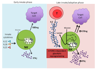
Figure 1. Summary of NK cell role in the innate immune response. Image taken from Gardiner & Finlay (2017). The major innate cytokines that activate NK cells are IL-12, IL-15, and IL-18 (11). NK cells carry out direct cytotoxicity of target cells and are potent producers of IFNγ (11). IL-2 is the cytokine that drives mTORC1-dependent glycolytic reprogramming of NK cells (11).

The authors on this paper worked with natural killer cells from humans as well as mice, which serve as valuable model organisms because their genetic, biological and behavioral characteristics resemble that of humans (3). Their first discovery was that the presence of excess fat clogs up the lytic machinery of NK cells by inducing lipid metabolism. That lipid accumulation affects their cellular energetics, resulting in “metabolic paralysis” (3), which is what leads to the eventual loss of anti-tumor function. In order to better understand the effects that obesity has, they examined mouse models of diet-induced obesity (3), performing transcriptional analysis to see how NK expression varied in mice with a high-fat versus a control diet (3). They found that the high fat diet led to a substantial upregulation of lipid handling and metabolizing genes in the NK cells. Those results suggest that obesity induces metabolic reprogramming in NK cells to lipid metabolism, which inhibits the expression of effector molecules that would be important in an innate immune response (3). Additionally, they found a significantly reduced number of high lipid content NK cells when comparing obese and lean individuals, which was consistent with previous studies (6). There was an accompanying loss of NK cell function in humans with presence of excess fat, as they had previously found in mice (3).
In examining the mechanisms underlying these alterations to NK function, researchers found that the mTOR (mammalian target of rapamycin) pathway was inhibited in obesity (3). This pathway is extremely important in regulation of the cell cycle (7). mTORC1 (the molecule of focus in this study) can be activated by diverse stimuli including growth factors, nutrients, energy and stress signals, and other essential signaling pathways (7). One of its main functions is to activate proteins that are important to RNA translation (7), though its most important role is as a regulator that controls cell growth, proliferation and survival (8). mTORC1 activation is crucial for NK cell function, particularly IFN-γ production (3). The researchers treated human NK cells with rapamycin, the main inhibitor of this pathway, and found that tumor-killing was greatly reduced in rapamycin-treated cells (3). Understanding the importance of the mTOR pathway in anti-tumor activity, they then examined mTOR’s response to lipid accumulation and the data indicated that mTORC1 activation was greatly reduced in obese individuals (3). Those findings confirm that inhibition of the mTOR pathway in response to lipid accumulation is a major mechanism for reduced tumor-suppressing activity of NK cells.
The study looked into another pathway important in NK cell function: the PPARα/δ pathway, which is linked to lipid metabolic pathways (9). The study found that PPAR target gene expression increased in mice and human obesity (3). Interestingly, they found that the PPAR pathway led to increased lipid uptake and inhibition of the previously mentioned mTOR pathway. When tested in mice, PPARα/δ agonists mimicked the effects of obesity on NK cell function (3).
One of the most interesting findings of the study was the reversibility of the metabolic defects in NK cells associated with excess fat (3). Researchers found that blocking Cpt1, a gene that is upregulated in obesity and involves transportation of fatty acids to the mitochondria, increased glycolysis by inducing a switch from oxidative phosphorylation (3). While both glycolysis and oxidative phosphorylation were defective in obesity, previous studies had demonstrated the importance of glycolysis in NK cell function (10). Therefore, they reasoned that increasing glycolysis would restore the cytotoxicity of NK cells (3). Both glycolysis increase and NK cell cytotoxicity was restored when they introduced the Cpt1 inhibitor etomoxir (3).
The researchers were able to mimic obesity through lipid administration and by using PPARα/δ agonists, which inhibit mechanistic target of rapamycin (mTOR)-mediated glycolysis (3). In other words, they found that the both those pathways were important to the NK cell defects in obesity. They used this to find that it was possible to reverse the harmful metabolic paralysis by (a) inhibiting PPARα/δ or blocking lipid transport (3). All of that suggests that metabolic reprogramming of NK cells could restore their anti-tumor activity in cases of human obesity (3), suggesting exciting new possibilities for antitumor therapies.
It is well documented that obesity is associated with immune dysregulation, but most studies have focused on other pathways associated with inflammation that interferes with insulin signaling and contributes to diabetes (2). This recently published study contributes knew knowledge of how obesity also impairs antitumor activity of the immune system.

References
1. Brigham and Women’s Hospital. (2017). Why the immune system fails to see cancer. Science Daily. From www.sciencedaily.com/releases/2017/06/170629142922.htm  
3. Michelet, X., Dyck, L., Hogan, A., Loftus, R. M., Duquette, D., Wei, K., O’Farrelly, C. (2018). Metabolic reprogramming of natural killer cells in obesity limits antitumor responses. Nature immunology,  https://www.nature.com/articles/s41590-018-0251-7.pdf
4. Calle, E. E., Thun, M. J. (2004). Obesity and cancer. Oncogene, 23: 6365-6378.
5. Vivier, E., et al. Functions of natural killer cells. (2008). Nature Immunology, 9: 503–510.
6. Lynch, L. A. et al. Are natural killer cells protecting the metabolically healthy
obese patient? Obesity (Silver Spring), 17: 601–605 (2009).
7. Pópulo, H., Lopes, J. M., & Soares, P. (2012). The mTOR signalling pathway in human cancer. International journal of molecular sciences, 13(2): 1886-1918.
8. Viel, S., Marçais, A., Guimaraes, F. S. F., Loftus, R., Rabilloud, J., Grau, M., Bienvenu, J. (2016). TGF-β inhibits the activation and functions of NK cells by repressing the mTOR pathway. Sci. Signal, 9(415): ra19-ra19.
9. Pawlak, M., Lefebvre, P., & Staels, B. (2015). Molecular mechanism of PPARα action and its impact on lipid metabolism, inflammation and fibrosis in non-alcoholic fatty liver disease. Journal of hepatology, 62(3): 720-733.
10. Gardiner, C. M., & Finlay, D. K. (2017). What fuels natural killers? Metabolism and NK cell responses. Frontiers in immunology, 8: 367.
11. Gardiner, C. M., & Finlay, D. K. (2017). What fuels natural killers? Metabolism and NK cell responses. Frontiers in immunology, 8: 367.

A New Subtype of Regulatory T Cell Functions in Intestinal Wound Healing


In reference to a new paper published in Nature Immunology by researchers at King's College London and the National Institutes of Health.
Despite what you might hear in advertisements, “boosting your immune health” isn’t always a good thing. An overactive immune response could contribute to developing an allergy or an autoimmune disease. Excess inflammation can also damage sensitive tissues that aren’t used to that sort of response. A key element in keeping the immune response from getting out of control is the regulatory T cell (Treg). These are a special subset of T cells, the adaptive immune cells that (along with B cells) allows the body to have stronger, faster immune responses to pathogens that it’s been exposed to in the past. This is accomplished by the manufacturing of memory B and T cells, which are produced during the first immune response and stick around for years after. Some early T cell progenitors will become cytotoxic T cells (CTLs) which kill diseased cells with . Others become helper T cells (Th), which help determine the type of immune response to use for any given infection. For instance, a Th1 mediated response will use CTLs and produce more inflammation than a Th2 response, which limits inflammation and prefers to use antibodies to fight the infection. Tregs, however, reduce the ability of CTLs and other cytotoxic immune cells to kill cells and cause inflammation.  Some Tregs are made in the thymus, while others differentiate from naïve T cells peripherally out in other tissues (Sakaguchi et al. 2008). Their anti-inflammatory action can be performed by the secretion of anti-inflammatory types of chemical messengers called cytokines, specifically TGF- ß and IL-10. Tregs can also interrupt the activation of other T cells by blocking B7, a cell surface marker which needs to bind other cells for the T cells to become activated.


Tregs either develop from T cell progenitors in the thymus or from naïve T cells in the peripheral tissues. They prevent other T cell variants from performing immune functions which may lead to autoimmunity, allergy, and other forms of harmful excessive inflammation (Sakaguchi and Powrie 2007).


In a new study by Povoleri et al., a new type of Treg was identified. This cell, dubbed a CD161+ T regulatory cell for the CD161 cell surface protein which distinguishes from other Tregs, has all the classical features of other Tregs. It expresses similar genes at similar levels, particularly the Treg-specific protein FoxP3 which is generally thought to designate T cells to differentiate into Tregs (Coffer and Burgering 2004). However, it has subtle differences in its T cell receptor as well as a slightly different combination of cell surface marker proteins. When the researchers compared CD161+ Tregs to naïve (not yet activated) and memory (primed and ready to stop inflammation) Tregs, they found the CD161+ Tregs were more effective in suppressing CTL activity. The CD161+ Tregs were also more useful in preventing the mouse immune system from rejecting tissue grafts, indicating that they suppress the inflammatory response more thoroughly than other Tregs. When each type of Treg was activated, the CD161+ Tregs produced more anti-inflammatory cytokines than either naïve or memory Tregs.

The researchers also found that the CD161+ Tregs are regulated by retinoic acid, a molecule that normally causes Tregs to develop in the intestines. Retinoic acid causes the CD161+ Tregs to express the CD161 protein as well as the CCR9 and ITGA4 genes, factors that cause cells to move to the intestinal tissues. In addition, the paper examined multiple protein binding sites on the CD161+ Treg DNA and found that binding of the protein BACH2 will decrease expression of many CD161+ Treg-specific genes. In this manner, BACH2 prevents the differentiation of CD161+ Tregs from T cell progenitors. Unsurprisingly, CD161+ Tregs show very low levels of BACH2 expression. Since other genes like CD161 that are regulated by BACH2 are often involved in wound healing, the authors theorized that CD161+ Tregs may play a role in healing wounds in the intestinal tissue that they localize to. 

These suspicions were confirmed when the authors found a higher abundance of CD161+ Tregs in the intestinal tissue than either naïve or memory Tregs. This trend was even stronger in individuals with Crohn’s Disease, a condition which causes excessive inflammation in the colon. It is likely that CD161+ Tregs are being trafficked to the inflamed tissues created by Crohn’s in order to reduce the inflammation. When wounded colorectal cells were grown in the presence of all three kinds of Tregs, the tissue healed most effectively when CD161+ Tregs were added. Taken together, this data all indicates that this new variation of regulatory T cell is vital for healing wounded tissue in the intestines. This could be incredibly useful new information in the fight against diseases like Crohn’s that cause severe pain from excessive inflammation. Perhaps other tissue-specific Treg subtypes exist that can be used to fight localized inflammation in different regions throughout the body.


References

Coffer, P. J., & Burgering, B. M. (2004). Forkhead-box transcription factors and their role in the immune system. Nature Reviews Immunology4(11), 889.

Sakaguchi, S., & Powrie, F. (2007). Emerging challenges in regulatory T cell function and biology. Science317(5838), 627-629.


Sakaguchi, S., Yamaguchi, T., Nomura, T., & Ono, M. (2008). Regulatory T cells and immune tolerance. Cell133(5), 775-787.