-->
Showing posts with label interferon. Show all posts

Monday, September 30, 2013

Correlation between HLA alleles and pH1N1 mortality rates


            Many people have had the flu at last once in their life and see their doctors every year for a flu vaccination.  The vaccine is typically trivalent, which means that it protects against three different strains of the influenza virus: H1N1, H3N2, and an influenza B strain.  This vaccine is given as an intradermal shot meaning it is injected into the skin instead of the muscle2.  Seeing as how flu season is fast approaching I though it fitting to bring to light an article relating the flu, or more specifically the pandemic influenza A virus strain (pH1N1) of the 2009-2010 flu season.  However, before delving into the article I would like to provide the basic, necessary background information.

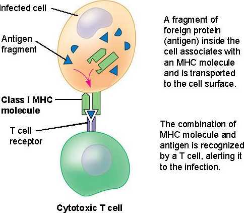
            When an antigen is engulfed by an antigen presenting cell (APC), its proteins are broken down and a small peptide chain of about 10 base pairs is expressed so that activated cytotoxic T-cells can destroy the infected cell3.  The proteins that express these peptides are called MHC (major histocompatabiltiy complex) class I and MHC class II proteins which are encoded by the human leukocyte antigen (HLA) gene3.  For MHC class I gene on HLA there are three regions: HLA-A, HLA-B, and HLA-C for which many different alleles exist3.  Some MHC alleles display the peptides for T-cell recognition better than others which is why different people have different immune responses to the same pathogen.  


Additionally, CD8+ is the specific receptor that MHC class I binds to on T-cells3.  Infected cells also release interferons (INFs) that increases the expression of MHC molecules which helps to fight off infection by causing more interactions with T-cells3.

Monday, September 16, 2013

Tiny Pests Causing Huge Problems


Why do people put on bug spray when they go outside?  The obvious answer is that having itchy mosquito bites all over your arms and legs is one of the most irritating feelings ever.  However, most people forget that these tiny insects can carry extremely dangerous viruses that can cause severe illness or even death.  West Nile virus (WNV), a well-known mosquito-borne virus, is the major cause of encephalitis (inflammation of the brain), which is a disease characterized by flu-like symptoms, and in severe cases, seizures and sensory/movement issues.  Dengue virus (DENV) is another dangerous mosquito-borne virus that is a major cause of viscerotropic disease, which is a disease that is associated with yellow fever and ultimately leads to organ failure and death.  These viruses cause significant problems all over the world, and currently there are no approved vaccines to prevent or combat WNV and DENV.  In order to develop effective vaccines, we need to understand how our bodies’ immune system recognizes the viruses and provide protection.  “Innate Immune Sensing of Flaviviruses” is a recently published paper in PLoS Pathogens, in which Suthar et al. (2013) discusses recent research findings on the way that the innate immune system interacts with WNV and DENV to trigger an antiviral response.1 Also, understanding how these viruses can evade our immune response is equally important for developing new treatments.  A group mentioned in “Innate Immune Sensing of Flaviviruses,” Aguirre et al. (2012)2, investigate one of the main mechanisms that DENV uses to inhibit inducing an immune response.
           
WNV and DENV are part of the flavivirus family, and they contain a single stranded RNA genome.  In order to recognize these viruses, we encode receptor proteins called pattern recognition receptors (PRRs), which recognize and bind to structures on pathogens that are not present in normal, human cells.  These structures are called pathogen-associated molecular patterns (PAMPs).  Once the virus is recognized, antiviral immune defenses are initiated which leads to inflammation.
  
The two PRRs that recognize the viral, non-self RNA structures are called retinoic-acid inducible gene-1 (RIG-1)-like receptors (RLRs) and myeloma differentiation factor 5 (MDA5).  RIG-1 recognizes and binds to double stranded RNA with a triphosphate group on the 5’ end.  MDA5 interacts with long double stranded RNA.  Double stranded RNA does not exist in normal, host cells, and it is a common viral PAMP.  Once the virus is recognized, RIG-1 and MDA5 induce the production of type 1 interferon (IFN) and proinflammatory cytokines that will migrate to the site of infection and recruit/help produce other factors needed to eradicate the virus.  A study done by Fredericksen et al. (2008) demonstrated that RIG-1 and MDA5 function cooperatively in creating an antiviral response to WNV.3 They found that RIG-1 is activated first by WNV, and then MDA5 is activated to sustain type 1 IFN and ISG expression (initiated by RIG-1).3   It is known that the PRRs RIG-1 and MDA5 are involved in pathogen recognition and initiate the immune response, but it is still unclear what the exact PAMP RNA substrate and nucleic acid sequence that RIG-1 and MDA5 interacts with. 

Friday, May 3, 2013

Interferon, Always Messing Stuff Up


Persistent viral infection such as those from HIV, Hepatitis B and Hepatitis C can lead to continuous pain and suffering in infected individuals. Persistent viruses are able to avoid clearance by down-regulating the immune system and suppressing the activation of antiviral CD4 and CD8 T cells. After persistent infection, a hyperimmune response is often observed characterized by elevated pro-inflammatory mediators and a constant interferon signature. Type 1 interferon (IFN-1) signaling is upstream of many immune responses such as activation of T cells, B cells and natural killer cells as well as inflammatory genes. Interferon is a protein that cells display during the presence of pathogens to communicate to other cells and initiate the innate immune system. Interferon signaling is necessary for host defense against viral infection and viral immunity during acute infections.(1) Because of this it is suspected that IFN-1 is responsible for the hyperimmune activity in persistent viral infection.

         To understand the role of IFN-1 in persistent viral infection the authors used the Armstrong (Arm) and clone 13 (Cl13) strains of lymphocytic choriomeningitis virus. The Arm strain is characterized by an acute infection, while the Cl13 strain is characterized by a systemic viral infection. Through the use of IFN-β-yellow fluorescent protein tagging in plasmacytoid dendritic cells (pDCs) it was determined that IFN-β was produced during Cl13 infection and not during Arm infection. Fluorescent protein tagging works by attaching an antibody to a cell surface protein receptor. Then an additional molecule that contains the fluorescent tag binds to the antibody attached to the protein of interest thereby allowing you to observe the expression of the target protein, in this case IFN-β .By replacing the glycoproteins of Arm and Cl13 viruses with green fluorescent protein, the authors confirmed that Cl13 infection preferentially targets pDCs. Green fluorescent protein is a protein that is found in jellyfish. We can insert the gene for the green fluorescent protein into the coding sequence of our target protein, then when the protein of interest gets transcribed the green fluorescent protein will be transcribed as well and we can measure target protein levels by measuring the level of green fluorescent protein. pDCs showed increased green fluorescent protein expression when infected with the Cl13 viruses marked with the protein compared to when infected with the Arm viruses marked with the green fluorescent protein.

Sunday, March 24, 2013

Tissue selectivity of West Nile virus infection


A recent study reveals insight into how West Nile virus is able to infect spleen but not liver tissue.

           West Nile virus (WNV) is most commonly transmitted to humans by mosquitoes that pickup the virus from infected birds. Originally identified in Africa in 1937, WNV is now a problem in the U.S since it spread to New York in the summer of 1999.3 Although infection isn’t always serious, when it is the virus can be very harmful. As a neurotropic virus it has the ability to infect nerve cells and the neurologic effects of infection can be permanent or deadly2,3,4. About 1 in 150 people will develop a severe illness as a result of the infection2. Since the initial outbreak in the U.S. the virus is still spreading around the country via mosquitoes2. To get an idea for numbers, about 30,000 people have been reported sick from WNV in U.S. since 1999.  Not every infection is severe because our bodies have mechanism to fight off the virus.
            Upon infection with West Nile virus, the virus is localized into particular tissues within our body, while other tissues remain “protected” from infection.  In humans, WNV infection is restricted to the skin, draining lymph node, spleen and central nervous system tissues like the brain and spinal cord. WNV does not infect the liver. We still haven’t quite figured out what controls whether WNV can infect one type of tissue over another. What are the differences between these tissues that cause a difference in the ability of WNV to infect? Are there cell factors present in the liver that restrict infection by WNV? These are the types of questions that researchers at University of Washington School of Medicine gave some answers to in a recent study published in February 2013.
            In order to address the question of what controls tissue tropism, we’ll we need to take a closer look at processes not visible by the naked eye. Previous research has established pathways inherent in our cells that are involved in fighting off virus infection. For instance there are receptors in our cells that recognize viral mRNA as foreign which initiates a response in order to fight off the virus. There are two major signaling pathways in our cells identified in previous research as important in this response. One is called RIG-1 and it recognizes viral RNA as a non-self pathogen. Once viral RNA is recognized, this eventually activates a secondary pathway involving proteins called interferons (IFN) that drive transcription to activate an anti-viral response.
             Research studies have found that there are some cases of infection, although rare, in peripheral organs like the liver1. Since WNV has the capability to infect liver cells in rare cases, this suggests that maybe those liver cells lacked the restriction factors normally present to block virus infection. Researchers at Washington School of Medicine investigated if the two pathways that are already well established by previous research to be involved in general restriction of virus infection (RIG-1 and IFN)5,6 also control what tissue types WNV can infect. They used mouse models to mimic WNV infection in humans. In both humans and mice, WNV infections are limited to the same tissues. They found that RIG-1 and IFN pathways did indeed regulate tissue specific responses to West Nile virus!
            So what’s different between spleen and liver cells that allows WNV to infect spleen cells? That’s exactly the question that researchers in this study were delving into. They found that if you block the RIG-1 and/or IFN pathways then upon exposure to WNV the liver cells become permissive to WNV. This indicated that these two signaling pathways (RIG-1 and IFN) are involved in restricting WNV from infecting certain tissue types. For infection of the spleen, which is a tissue that usually gets infected by WNV, it took about 4 days for the amount of virus to build up to significant levels. Once the RIG-1 and/or IFN pathways were blocked, the virus was able to infect spleen cells much earlier suggesting that these pathways have a function in spleen cells to restrict early stages of viral replication.
            Now these pathways have many genes involved with them, so the network of what is going on is very complex. Therefore the researchers took a systems biology approach in order to investigate what genes are being differentially expressed in spleen versus the liver tissue that impacts WNV infectivity. They were able to categorize hundreds of genes which were differentially expressed between spleen and liver tissue in response to viral infection. Their systems biology approach confirmed previous findings of restriction factors that block WNV virus infection, and also found new ones! The new genes identified can now be  investigated for furthur research. Once we get a better handle on what is going on in our bodies to fight off West Nile virus, we will have a better chance at stopping virus infection. So until further research, watch out for those mosquitoes!

Primary Article:

1) Suthar MS, Brassil MM, Blahnik G, McMillan A, Ramos HJ, Proll SC, Belisle SE, Katze MG, Gale M (2013) A Systems Biology Approach Reveals that Tissue Tropism to West Nile Virus is regulated by Antiviral Genes and Innate Immune Cellular Processes. PLOS pathogens. Vol 9:2.

Supplementary Sources:
  

3) http://www.cdc.gov/ncidod/dvbid/westnile/index.htm
4) Samuel MA,  Diamond MS (2006) Pathogenesis of West Nile Virus Infection: a Balance between Virulence, Innate and Adaptive Immunity, and Viral Evasion. J Virol. 80: 9349-9360.
5) Ramos HJ, Gale  M, Jr. (2011) RIG-1 like receptors and their signaling crosstalk in the regulation of antiviral immunity. Curr Opin Virol 1: 167-176. Schoggins JW, Wilson SJ, 6) Panis M, Murphy MY, Jones CT, Bieniasz P, Rice CM ( 2011). A diverse range of gene products are effectors of the type I interferon antiviral response. Nature. 472: 481-485.
Monday, February 25, 2013

Fighting Fire with Fire


To some it may seem counterintuitive to use an infection to cure a disease. Surprisingly enough, this is exactly what researchers have been doing using oncolytic virus therapy.  Oncolytic viruses are naturally occurring or engineered viruses that selectively target cancer cells by identifying common cancer characteristics such as defective immune responses or abnormalities in cellular signaling pathways. Once viruses infect the cancer cells, they replicate within the cell and eventually kill it.  As such, the infectious qualities of oncolytic viruses are becoming increasingly useful tools during cancer treatment. 


Past studies have shown great potential for oncolytic therapy using vesicular stomatitis virus, (VSV) -- a negative-strand RNA virus that generally infects insects, cattle, horses and pigs (1).  VSV has proven to be particularly conducive to oncolytic virus therapy as a result of its small easily manipulated genome and relative innocuity in humans (2). So far, VSV vectors have been used for immunization against HIV and influenza (2,3).  Additionally, testing has occurred that suggests that there is also potential of VSV as a recombinant cancer vaccine vector (4). 


Despite the high hopes for the use of VSV as oncolytic cancer treatment, a recent 2012 study suggests that human pancreatic ductal adenocarcinoma (PDA) cells do not consistently permit VSV infection (5).  In some cases, PDA cells retained all resistance and were still able to activate functional interferon (IFN) responses. As a result, the study of the interferons – proteins released by host cells to interrupt viral replication – and their association with oncolytic VSV infection is of great interest to researchers.

Wednesday, January 4, 2012

Like a finely fitted suit: IKKε tailors the antiviral response.


Every pathogen is different, so every immune response must be different. There is no “one size fits all” garment in immunology! This tailoring can occur at multiple levels. One systemic response is the development of “Th1” versus “Th2” CD4 T cells, which facilitate immune responses against (generally) intracellular and extracellular pathogens, respectively. On a more localized scale, different cell types, such as cardiac fibroblasts and myocytes, express different basal levels of immune components to dictate how the immune response behaves in particular tissues, such as the heart (1). Recent research by Sze-Ling Ng and colleagues has highlighted another level of tailoring, at the level of how individual cells coordinate their gene expression in response to distinct antiviral cytokines.
            Interferons are central components of the antiviral response. There are several types of these secreted proteins, including type I interferons (IFN-I), which help cells inhibit virus replication, and type II interferons (IFN-II), which are pro-inflammatory: they recruit immune cells to the site of infection. Both types act in a similar manner, by binding to receptors on the cell surface and inducing signals that culminate in a change in gene expression in the cell. Although IFN-I and IFN-II bind to different receptors, they share components of their signal transduction mechanism, including the transcription factor STAT1. Following IFN-I signaling, STAT1 becomes active and binds with two other proteins, STAT2 and IRF-9. Together, this complex binds to specific DNA sequences to mediate transcription of genes (interferon-stimulated genes, or ISGs) that mediate the antiviral state. Following IFN-II signaling, STAT1 also becomes active, but binds to itself to form a STAT1:STAT1 dimer. The STAT1 homodimer binds to DNA to mediate transcription of a distinct, but partially overlapping, set of genes that lead to the inflammatory response. A central question has been how cells balance these two responses, especially since both IFN-I and IFN-II are likely to be produced upon pathogen infection. Ng et al. now demonstrate that a cellular kinase, IKKε, phosphorylates STAT1, to alter its transcriptional response to IFNs.
            IKKε is best known for its role in inducing production of IFN-I in response to viral infection. However, genetic studies in mice have shown that a highly related kinase, TBK-1, is sufficient to induce IFN-I, raising the question of how IKKε contributes to the interferon response. One possibility is that IKKε is only required in a subset of specialized cells. Alternatively, because IKKε is inducible whereas TBK-1 is constitutively present, others speculated that IKKε functioned as a positive feedback mechanism. It was previously shown that IKK<ε could phosphorylate STAT1 on a serine at position 708 (2), and that this modification was necessary for an efficient immune response. Ng et al now show that this phosphorylation alters the capacity of STAT1 to form homodimers, leading to increased formation of the STAT1:STAT2:IRF-9 complex, and altering the balance of IFN-I vs. IFN-II transcription in cells.

The discovery of a new class of memory CD4 T cells in the lung may lead to more effective flu vaccines


                  In my experience, the close of the semester and the beginning of the holiday season generally coincides with a rapid collapse into illness brought on by lack of sleep, constant stress, poor nutrition and far too much time spent in the library away from natural light.  Unfortunately, many times this mental and physical collapse corresponds with an actual illness: the flu.  Even though I was one of the first on campus to get a flu shot (did you get yours?), I could still possibly contract the flu in addition to the aforementioned collapse, as each year the vaccine producers guess which strain of flu will be most likely to spread.  If they miscalculated and selected the wrong strain, then my exhausted classmates and I
could be in for a fun-filled break.  
                  A recent study uniting scientists from the
University of Maryland School of Medicine, Columbia University, the University of Connecticut Health Center and the University of Pennsylvania School of Medicine identified a new class of memory CD4 T cells, in the lungs; research based on this discovery could eventually lead to a flu vaccine that would remove the possibility of my classmates and I needing to cope with the flu on top of exhaustion and stress induced illnesses.  Memory cells are produced following initial exposure to a pathogen and “remember” specific components of the pathogen, in case a person is infected with the same, or highly similar, pathogen in the future. CD4 T cells, also called “helper” T cells, coordinate the adaptive immune response, by assisting B cells to produce antibodies and CD8 T cells to kill infected cells. This study utilized the identification of influenza-specific, stationary, memory CD4 T cells in the lung to examine their memory in comparison with CD4 T cells found in the spleen.  This experiment was carried out with mice that were infected with a specific strain of influenza; the mice who had memory CD4 cells in their lungs only did not die, whereas the mice with CD4 T cells in their spleens seemed to have a greater likelihood of dying from the influenza, even though their T cells were found in a more diverse area and had the ability to migrate to peripheral tissue sites to encounter the infection.
Generally, if T cells have the chance to migrate to the peripheral tissues to directly engage the infection, then they will be more effective at attacking and removing the pathogen.  The memory T cells produced can then remain in the tissues which had been infected or they can move to other areas of the body until they are called into action upon the return of the pathogen.  In order to design a vaccine that can effectively target the most common tissue infection site for a specific pathogen, it must be known which location for the memory T cells is most effective. 
The hypothesis tested in this study was that the specific tissue site of the CD4 T cells would play a key role in determining their homing abilities and thus their success at protecting the infected mouse from dying from the influenza strain used in the experiment, H1N1.  The lung memory CD4 T cells were found to produce a greater amount of IFN-gamma, an important inflammatory messanger, than the spleen memory CD4 T cells, while the spleen memory CD4 T cells had a greater amount of IL-2 producers than the lung memory CD4 cells.  These values show that resident memory populations may function independently from the specifics of the antigen.  The lung memory CD4 T cells remained in the lung and contain so called “zip codes” which let them identify the tissue they are supposed to be in and concentrate their efforts specifically in the site of the infection; the spleen memory CD4 T cells do not have these zip codes.  An additional comparison between lung memory CD4 T cells and lung memory CD8 T cells revealed the special zip code feature of the CD4 T cells, as the CD8 cells may migrate to lymph tissues and non-lymph tissues.  Because of the homing device, the lung memory CD4 T cells have increased protective abilities; all the mice containing just this type of T cell survived, whereas the spleen mice, whose CD4 T cells could migrate to other tissues, were not as fortunate. 
This newly discovered CD4 T cell offers wonderful protection against respiratory infections, including influenza.  Their protective capabilities are due to their “zip code” homing behavior, placing them directly in the site of the infection.  Further study proposed by these researchers would be to examine how knowledge of compartmentalized CD4 T cells can be applied to vaccines.   If a vaccine can be made to target a specific tissue and generate specific memory T cell that will remain in that tissue, then it will be far easier to combat specific strains of antigens.  This would be especially useful for influenza.  Hopefully this will become a reality and the flu shot will be guaranteed to prevent the flu (though students may still have to deal with the end of semester collapse).

Teijaro, J. R., Turner, D., Pham, Q., Wherry, E. J., Lefrancois, L., & Farber, D. L. (2011). Cutting edge: tissue-retentive lung memory CD3 T cells mediate optimal protection to respiratory virus infection. The Journal of Immunology, 187, 5510-5514.

Post by Jessie Solcz

Saturday, December 24, 2011

Hey Virus: Replicate Here!


The innate immune system, that part of the immune system that responds to non-specific signs of pathogen infection, faces a quandary when mounting a response against a pathogen. On one hand, it must limit the replication of the pathogen to prevent spread throughout the body. On the other, it must facilitate the development of a specific adaptive immune response, which can clear the pathogen from the body and lead to immunologic memory. It might seem like both of these are reasonable tasks, but the quandary lies in the fact that the adaptive immune system must be activated by a certain amount of antigen (proteins derived from the pathogen that can be effectively targeted by the immune system) in order to elicit an effective response. Low levels of antigen lead to weak responses, whereas high amounts of antigen induce strong responses. Thus, if the innate immune system does the first part of its job too well and largely prevents virus replication, there will not be enough viral antigen present to stimulate a robust adaptive response. So how does the immune system get around this predicament? A new paper in Nature Immunology by Nadine Honke and her colleagues sheds some light on this question (1).
            The major weapons in the innate immune system’s arsenal are small, secreted proteins called interferons. When cells in the body detect a pathogen, they produce interferons, which alert the surrounding cells to the impending danger. These “warning flags” bind to interferon receptors found on the surface of all cells, and induce the expression of a large number of genes, termed interferon stimulated genes (ISGs), which can combat the pathogen through various means. Interferons also initiate mechanisms that facilitate the adaptive immune response, by increasing the presentation of specific antigens to the T cells and B cells that comprise the adaptive immune system. To effectively stimulate T cells, however, requires a sufficient dose of antigen: prior studies have indicated that over 20,000 copies of the antigen are required for an antigen-presenting cell to induce a robust T cell response (2). So, how can the innate immune system effectively prevent the spread of a pathogen while still providing enough “grist” to be presented to naïve T cells? Honke et al began to answer this by examining a particular subset of innate immune cells, metallophilic macrophages.

Friday, December 16, 2011

New EAE Models More Accurately Reflect MS

Multiple Sclerosis (MS) affects about 400,000 people in the United States. In MS, the myelin sheath that coats our neuronal axons is degraded, as are the cells that produce myelin (also known as oligodendrocytes). MS is considered an autoimmune disease because the attack is facilitated by our body’s own immune system. This degradation of the myelin sheath affects the ability of our neurons to transmit electrical signals to each other. This manifests itself in the symptoms often associated with MS: numbness in limbs, paralysis and vision impairment.

In order to study MS, researchers often employ the use of animal models. Specifically, Experimental Autoimmune Encephalomyelitis (EAE) is a well recognized mice model that mimics the progression of MS. EAE is considered a Th1 focused disease with T cells secreting primarily IFNϒ. T cells are immune cells in the body that participate in cell-mediated killing of foreign pathogens (1). In MS, they recognize our myelin as a foreign substance and proceed to destroy it. One way they do this is by secreting cytotoxic cytokines, such as the aforementioned IFNϒ. When inducing EAE in mice, this Th1 response is ensured by injecting a myelin peptide(to mount an immune response against) along with complete Freunds adjuvant (CFA), which contains a bacterium called M. Tuberculosis (CFA).

Aside from IFNϒ, IL-23 has emerged as a notable cytokine because mice deficient for it remained protected against EAE pathology. Furthermore, IL-23 promotes the differentiation of inflammatory Th17 cells (2). Numerous EAE models currently exist; some more representative of MS in certain clinical regards (e.g., onset, clinical progression, and remission). Therefore, it’s vital to always explore new EAE models in an effort to find one that best represents human MS. In a recent study by Smith et al. 2011, researchers replaced M. tuberculosis with C. rodentium (CRA)in the injected adjuvant. CRA is a bacteria known to induce an IL-23 dependent Th17 response (as opposed to the aforementioned M.tuberculosis-mediated Th1 response) to find out whether different EAE phenotypes would emerge.

CD4+ T cells in HIV

Human immunodeficiency virus (HIV) is a member of the retrovirus family and progresses into AIDs after a period of time. AIDs is defined by a significant deficiency in T-cell count which causes a progressive failure of the immune system and allows life-threatening opportunistic infections and cancers to thrive. Infection with HIV occurs by the transfer of blood, semen, vaginal fluid, or breast milk. In order to slow the progression to AIDs, many people with HIV take antiretroviral therapy which combines a number of drugs that are designed to stop HIV from infecting cells: includes nonnucleoside reverse transcriptase inhibitors (NNRTIs), protease inhibitors, and triple-nucleoside (or nucleotide) reverse transcriptase inhibitors (NRTIs) (1). HIV initially depletes the body’s CD4 T-cells and the point of this therapy is to boost the immune system back to normal to continue to fight the infection. However, 30% of patients with HIV who receive antiretroviral therapy fail to achieve a normal CD4+ T-cell count (2). This failure to achieve normal T-cell levels results in a steady decrease in T-cells to where, in less than 10 years, the count is below 500 (signifying AIDs) leaving the patient open to opportunistic infections and eventually death. Therefore it is necessary to study the reasons for this initial failure to reach normal T-cell counts with antiretroviral therapy in order to enhance its function.
HIV first infects CD4 T-cells, depleting the amount in the body. However, there is normally a period of time in which the T-cells regenerate in order to fight the infection. The balance of T-cells in the body is negatively regulated by interferon α (IFN-α) and evidence suggests that HIV increase the production of this interferon (3). This regulating interferon may promote apoptosis of uninfected CD4+ T cells by up-regulating expression of a death signal (TRAIL) and its death receptor (TRAIL receptor) (4). Therefore, the role of IFN-α may have important effects on CD4 T-cell populations in HIV patients.

A study by Sonia Fernandez at the School of Pathology and Laboratory Medicine in Australia looks at the effects of IFN-α on CD4 T-cell populations in HIV patients. They recruited HIV patients who have been receiving effective doses of antiretroviral therapy and measured the levels of CD 4 T-cells in the blood as well as levels of IFN-α. The patients were divided into low or high CD4 T-cell groups. Expression of markers of T-cell activation (HLA-DR), apoptotic potential (Fas), or aging (CD57) were assessed and it was found that the proportions of CD4+ T cells expressing HLA-DR or Fas were higher in patients with low CD4+ T-cell counts than in those with high counts such that the higher the levels of Fas and HLA-DR that are expressed, the lower the population of CD4+ T-cell count. The proportion of CD4+ T cells expressing CD57 did not differ between patients with low or high CD4+ T-cell counts. They left the cells in a culture for 72 hours and found that the proportions of CD4+ and CD8+ T cells that were apoptotic or preapoptotic was similar in patients with low and high CD4+ T-cell counts.

Wednesday, December 14, 2011

Immunotherapy: Giving Our Bodies a Boost to Fight Infection

The human body’s immune system works tirelessly to keep us healthy. So wouldn’t it be nice if we could lend it a hand in its effort to rid our bodies of all types of pathogens and cancers? Immunotherapy is a type of treatment that seeks to augment our own body’s immune system as it battles certain illnesses and physical conditions. Applications of immunotherapy have been found to be significant in the treatment of cancer, AIDS, autoimmune diseases, and in protecting the body following organ transplantations.

Immunotherapy can be broken down into two main categories: active immunotherapy and passive immunotherapy. Active immunotherapy is designed to stimulate our own immune system so that it can be more effective in combating a disease, such as a cancer vaccine. On the other hand, a passive immunotherapy uses artificially produced components of the immune system, such as antibodies, as a form of treatment. Playing an important role in immunotherapy are certain molecules known as immunomodulators. These molecules, including IL-2, IL-7, IL-12 and interferon, interact with the immune system to induce, enhance, or even suppress the actions of the immune system. More specifically, IL-2 can enhance T, B, and NK cell activation, while IL-7 can promote the development of lymphoid progenitor cells from hematopoietic stem cells. IL-12 is important for the differentiation of ThO cells from naïve T cells and interferon improves antiviral responses. In order to develop new kinds of immunotherapy, researchers must first acquire a sound understanding of how the immune system functions as well as the nature of the particular illness being confronted.

Friday, December 9, 2011

Don't forget the bug spray! Establishing T-cell and memory T-cell responses to malaria


Malaria, and its causative agent, the parasite Plasmodium falciparum, is transmitted person-to-person through a bite from an Anopheles mosquito.  After being bitten by an infected mosquito, P. falciparum parasites in the sporozoite stage (PfSpz) enter the body and travel to the liver where they infect liver cells—this stage of the disease is asymptomatic.  In liver cells the sporozoites divide into merozoites (PfRBC) that then exit the liver cell and infect red blood cells.  This infection of red blood cells causes the symptomatic stage of malaria.
In the field of public health, it is generally accepted that immunity from malaria infections is usually short-lived and weak at best.  A study by Teirlinck et al published December 1 in PLoS Pathogens set out to test whether or not lasting immunity is actually induced by infection with P. falciparum.  In order to direct their experiment, the researchers chose to investigate recent findings that cytokines, small signaling proteins, play a role in the cellular immune response to P. falciparum.  To do this, the researchers infected two groups via mosquito bites; group A was treated with the prophylactic anti-malaria drug chloroquine while group B received no prophylaxis.

Monday, December 5, 2011

Interfering with Pancreatic Cancer Growth – Role of Interferon Regulatory Factor-2 in Pancreatic Tumorigenesis


Apple CEO Steve Jobs; 2011 Nobel Prize Laureate Ralph Steinman; actor Patrick Swayze; actor Michael Landon; opera singer Luciano Pavarotti; comedian Jack Benny; actress Donna Reed; musical composer Henry Mancini. These well-known individuals, despite varying career paths, share a rather tragic connection: each has lost his or her life as a result of pancreatic cancer. Pancreatic cancer, or pancreatic carcinoma, has the highest mortality rate amongst all major cancers. Within 5 years of diagnosis, approximately 94% of patients will die from this form of cancer. Just this year, it is estimated that 44,030 cases of pancreatic cancer will be newly diagnosed, while approximately 37,660 patients will have died as a result of the disease [2]. Cigarette smoking, obesity, type II diabetes, blood types A/B/AB, and loosely defined genetic influences may present elevated risk for developing pancreatic carcinomas; however, the majority of patients diagnosed with pancreatic cancer do not exhibit any identifiable risk factors. Pancreatic cancer is a rather aggressive malignancy, and the high mortality rate is attributed to the difficulty in detecting the cancer at an early stage, as well as to the complexity involved in its development. Because this form of cancer has frequently reached advanced stages by the time of diagnosis, removing the tumor through surgery is not often a treatment option. When the tumor has spread beyond the pancreas (metastasized) and is not operable, chemotherapy alone, or in combination with radiation therapy, is utilized. Traditionally, the chemotherapy drug selected for treatment is gemcitabine [3]. Such limited and largely unsuccessful therapeutic options available for pancreatic cancer patients underscores the importance of defining new mechanisms for treatment.

Thursday, December 1, 2011

Developing a malaria vaccine: via IV or not via IV, that is the question


Malaria, the veritable scourge of Africa, causes almost 1 million deaths and 300 million sicknesses yearly, and is caused by a parasite that infects liver cells, or hepatocytes, and red blood cells.  This kind of parasite is known as an intracellular parasite—in the case of malaria, the most fatal causative agent is Plasmodium falciparum.  P. falciparum is transmitted by a mosquito bite, usually from an Anopheles mosquito. P. falciparum has development cycles that occur in the gut of the mosquito, and the liver and red blood cells of humans: because of this incredibly complex lifecycle, P. falciparum is very difficult to develop a vaccine.  In the past, vaccines that contain proteins from the surface of the parasite, but not the parasite itself—known as subunit vaccines—have been developed, but did not give high-level immunity (Draper et al 2010).  As a result, the development of a vaccine that contains the entire live parasite in a non-infectious form has been explored.  This type of vaccine, known as a live attenuated vaccine, has been manufactured—the results of the first human clinical trials are explained in a paper by Epstein et al published in Science Magazine.
The researchers developed a vaccine that contained live attenuated, non-reproductive sporozoites, the infective stage (but not the symptomatic stage) of P. falciparum.  The researchers wanted to test the effectiveness of the vaccine based on different transmission routes.  The transmission routes tested were: intra-dermal (ID)—in the skin—and sub-cutaneous (SC), or just below the skin.  These two routes were tested because of their similarity to the natural mode of transmission of the parasite.  A mosquito bite does not penetrate down to the muscle; instead the proboscis (the part that the mosquito sucks blood with) is inserted ID, and the researchers attempted to mimic this transmission.
 Differing concentrations of the live attenuated parasite were administered to three different groups once a month for four months.  Another group received the same treatment, but also received two boosters three and four months after the initial treatment.  After administering the vaccine, Epstein et al tested blood smears from patients every two weeks in order to ensure that the vaccine was properly attenuated and would not develop into the symptomatic stage of malaria, known as merozoites.  While none of the vaccines developed into merozoites, half the patients suffered from an “adverse event:” headache, malaise, fever, or aches.  While these data suggest that the vaccine may be bad because half of the patients had adverse reactions, something to keep in mind is that flu vaccines, tetanus boosters, and myriad of other vaccines cause similar “adverse events” in similar numbers of patients.

Friday, November 4, 2011

IRF-1 is Essential for Immunity Against West Nile Virus Infection in Mice

The West Nile Virus (WNV) is spread via mosquitoes and has been affecting humans in the United States since 1999 (1). Current research by immunologist further dissects antiviral immunity, in hopes of understanding WNV infectivity and how it can be combated or prevented. Previous research in mice has identified several immune mechanisms of control against WNV including cytokines, chemokines, complement, B CD4+ and CD8+ T cells (reviewed in [1]). These are all various aspects of the innate and adaptive immune system that aid in fighting against pathogens. More specifically, type 1 IFN (IFN-αβ) has been given special attention, as mice deficient in it, rapidly succumb to WNV infection (2). Further, it was determined that IFN induction is dependent on certain transcriptional signals. Past research pointed Brien et al. (2011) in the direction of IRF-1, as it has been reported to contribute to IFN-β induction. For example, it was found that IRF-1 transcription factor activated IFN-β gene transcription and regulated genes that directly impeded replication of several viruses. Further roles of IRF-1 have been found in controlling herpes virus, even though mice lacking IRF-1 did not have defects in their type I IFN response. This would suggest that IRF-1 regulates IFN genes “in a pathogen and cell type-dependent manner” (1). Further research has also identified IRF-1 as a tumor suppressing gene and a regulator of the adaptive immune response (3).
To understand the role of IRF-1, Brien et al. (2011) assessed WNF infectivity in IRF-1 normal and IRF-1 deficient mice (1). As previously mentioned, they chose to manipulate IRF-1 because mice lacking it (IRF-1-/- ) were vulnerable to WNV infection. First, it was confirmed that IRF-1 is required for control of lethal WNV infection. After infection with WNV, Wild-type (WT) mice had a 65% survival rate and a mean time to death of 11 days, whereas IRF-1-/- mice had a 0% survival rate and a mean time to death of 9.5 days.
To better understand how IRF-1 deficiency is a disadvantage for mice with WNV infection, the ‘viral burden’ was measured at various points post infection in serum, several peripheral organs, and the central nervous system (CNS). Indeed, increased levels of viral RNA were found in the serum and lymph nodes in IRF-1-/- mice compared to the WT. Therefore, IRF-1 controls the early stages of WNV infection. Additionally, WNV infected the spleen more rapidly and clearance was delayed in IRF-1-/- mice. WNV was also detected sooner in the kidneys of IRF-1-/- mice, further suggesting that IRF-1 normally functions to control infection in peripheral tissues.