-->

Friday, January 13, 2012

Eosinophils and asthma in mice

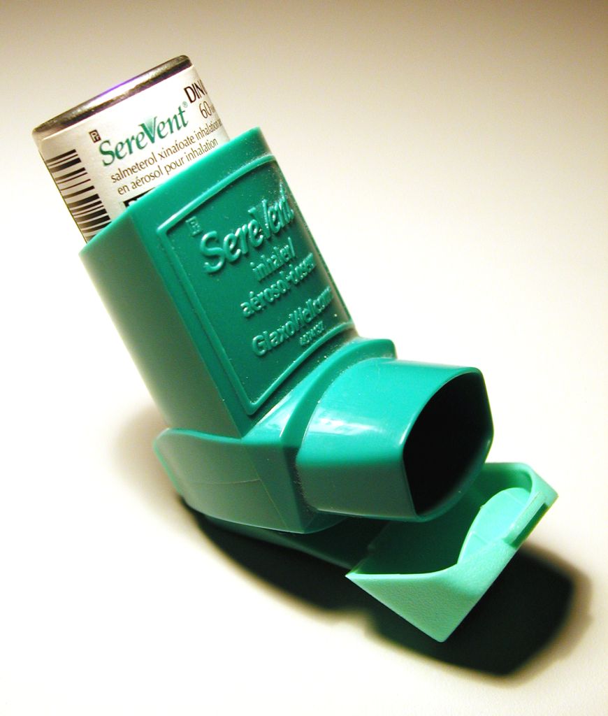
      Asthma is a disease that most Americans have knowledge of from childhood.  In grade school, there were always one or two children in class who either had to have their inhaler at the ready during recess and gym or were unable to take part in playground antics in fear of becoming short of breath.  As years passed, it became less obvious who was afflicted with asthma; as assignments would pile up, recess went away.  Yet, asthma remained.  It is not just an illness of childhood, it can affect people of any age, ethnicity and occupation.  It can be caused in part by genetic factors or by environmental triggers, especially proximity to smokers or excessively smoky areas.  This chronic disease emerges from the adaptive immune system’s response to “environmental assaults” and leads to constriction of the airway, coughing, tightness in the chest and shortness of breath.  The use of an inhaler with a corticosteroid will open the airway back up and allow the afflicted to breathe easy…at least until their next attack.  In order to better combat asthma, it is necessary to understand the mechanisms that make it so dangerous
      Asthma is a “type I hypersensitivity reaction,” meaning that it is caused by the secretion of a particular type of antibody, IgE, following a Th2 immune response. Th2 responses are one of the major types of immunity, and target extracellular pathogens. The Th2 class of helper T cells secrete cytokines that stimulate B cells to produce IgE, which can activate other types of cells to respond to antigens. When these IgE antibodies are targeted against environmental allergens, and not pathogens, type I hypersensitivity results, and when the response occurs in the bronchioles, it results in asthma. A recent study at the Mayo Clinic Arizona on asthma examined the role of eosinophils, a type of specialized white blood cell, in the regulation of dendritic cells and Th2 pulmonary immune responses with T cells in asthmatic mice.  There have been rumblings that the current description of eosinophils as “end-stage destructive effector cells” is not broad enough and that they in fact are involved with the secondary immune responses which lead to the activation and proliferation of memory T cells.  The DCs present the antigen to the lungs; in studies of asthma in mice, including this one, the mice were given additional DCs to better spread the allergen triggering antigen.  If a mouse has fewer eosinophils, then they generally will have reduced Th2 pulmonary abilities.   This study suggests that in allergen-specific T cell responses, eosinophils and DCs work together and are equally important and lead to a Th2 polarized immune response.  The eosinophils were found to have a profound impact on whether the inflammation of the respiratory tract goes by a Th2 pathway or a Th17 (a different “flavor” of immune response) pathway. 
      The relationship between the location of the eosinophils and their abundance, as compared with the accumulation of T cells during an asthma attack, was studied with three types of mice: eosinophil-null (PHIL mice), eosinophil-sufficient (wild type) and eosinophil-low (IL-5-/- mice).  PHIL mice had absolutely no eosinophils in their system, while the IL-5-/- mice had a partial loss of eosinophils compared to the wild type.  The mice were all treated with injections of an innocuous allergen under conditions that trigger asthma (OVA/Alum) and were subsequently exposed to airborne antigens.  The effects of the airborne antigens on the mice were then examined.  The comparisons between the three types of mice reveal information about whether or not the mice were able to accumulate DCs and eosinophils.  The draining lymph nodes of the PHIL mice did not acquire DCs in the 20 hours after they were exposed to the airborne antigens. In contrast, within 20 hours the nodes of the wild type mice acquired both eosinophils and MHC II activated DCs.  The researchers concluded that eosinophils are necessary for DCs to move to the lymph nodes and to cause T cell activation.  The mice that lacked the proper amount of eosinophils could not have the correct Th2 polarization in their lungs once they were exposed to an antigen, as the T cells were not properly activated in the LDLNs. 
      There is certainly more to be learned about eosinophils, DCs, T cells and their role in causing asthma.  The researchers suggest that eosinophils as monitors of localized immune responses can provide wonderful insights into the wider role of eosinophils in the immune system, as their impact on inflammation must certainly involve more than just allergies and asthma. 

Jacobsen, E. A., Zellner, K. R., Colbert, D., Lee, N. A., & Lee, J. J. (2011). Eosinophils regulate dendritic cells and Th2 pulmonary immune responses following allergen provocation. The Journal of Immunology, 187, 6059-6068.

Post by Jessie Solcz

Monday, January 9, 2012

Keep the hygiene, lose the inflammation: TGF-β and helminthic therapy

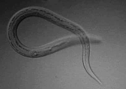
            Incidences of autoimmune and autoinflammatory diseases, such as type 1 diabetes, rheumatoid arthritis, and inflammatory bowel disease, are increasing in the developed world, and the annual costs of treatment amount to billions of dollars in the United States alone (1). There are numerous factors that can account for this rise in prevalence, including genetic differences and environmental factors. One factor that has been gaining support, both correlatively and experimentally, is the hygiene hypothesis: basically, since we have eliminated many of the prominent childhood diseases, children are “too clean,” and thus their immune systems, rather than focusing on pathogens, are stimulated to attack “self” targets instead, or target innocuous factors leading to allergy. In other words, infection with some types of pathogens, particularly parasitic worms such as roundworms, flatworms, and hookworms, can dampen the inflammatory responses that underlie many of these autoimmune/autoinflammatory conditions. We have largely eliminated these infections, which are transmitted through unclean drinking water and soil, in developed nations, thus accounting for some of the rise in autoinflammatory diseases.
            Some suffering patients have taken this idea to the extreme, and have deliberately infected themselves with parasites to try to alleviate their symptoms. Although this treatment is highly experimental and there is not a lot of clinical data demonstrating efficacy, anecdotal evidence suggests that some patients have observed marked reduction in symptoms following “helminthic therapy.” Obviously, there are many problems associated with introducing parasitic organisms into people, so if scientists can determine how helminthic infection tamps down inflammatory responses, it could potentially lead to novel anti-inflammatory treatments that don’t involve the parasites themselves.
            At the core of this issue are two major categories of immune responses, named “Th1” and “Th2” responses for the types of helper T cells that facilitate them. Th1 responses are inflammatory in nature, and involve the activation of macrophages and killer (CD8+) T cells. These responses are elicited by the secreted cytokine interferon-γ (IFN-γ), and typically target intracellular pathogens such as viruses. Th2 responses are mediated largely by B cells, which make antibodies, and are elicited by the cytokine interleukin-4 (IL-4). Th2 responses target extracellular pathogens, such as parasitic worms, and the rationale behind the hygiene hypothesis is that by eliminating many of these Th2 pathogens, the immune system “skews” towards Th1 responses. This, then, leads to increased inflammation and the associated autoinflammatory diseases. However, other responses caused by helminth infection, including the generation of regulatory T cells (TRegs) and the secretion of immunomodulatory cytokines such as IL-10 and transforming growth factor-β (TGF-β), might also mitigate autoinflammatory disease. A recent paper by Hübner et al attempted to distinguish which of these mechanisms was responsible for the protection from autoimmunity accorded by helminth infection. They found that generation of a Th2 response was not required, but that the production of TGF-β was largely responsible for protection.

Wednesday, January 4, 2012

Like a finely fitted suit: IKKε tailors the antiviral response.


Every pathogen is different, so every immune response must be different. There is no “one size fits all” garment in immunology! This tailoring can occur at multiple levels. One systemic response is the development of “Th1” versus “Th2” CD4 T cells, which facilitate immune responses against (generally) intracellular and extracellular pathogens, respectively. On a more localized scale, different cell types, such as cardiac fibroblasts and myocytes, express different basal levels of immune components to dictate how the immune response behaves in particular tissues, such as the heart (1). Recent research by Sze-Ling Ng and colleagues has highlighted another level of tailoring, at the level of how individual cells coordinate their gene expression in response to distinct antiviral cytokines.
            Interferons are central components of the antiviral response. There are several types of these secreted proteins, including type I interferons (IFN-I), which help cells inhibit virus replication, and type II interferons (IFN-II), which are pro-inflammatory: they recruit immune cells to the site of infection. Both types act in a similar manner, by binding to receptors on the cell surface and inducing signals that culminate in a change in gene expression in the cell. Although IFN-I and IFN-II bind to different receptors, they share components of their signal transduction mechanism, including the transcription factor STAT1. Following IFN-I signaling, STAT1 becomes active and binds with two other proteins, STAT2 and IRF-9. Together, this complex binds to specific DNA sequences to mediate transcription of genes (interferon-stimulated genes, or ISGs) that mediate the antiviral state. Following IFN-II signaling, STAT1 also becomes active, but binds to itself to form a STAT1:STAT1 dimer. The STAT1 homodimer binds to DNA to mediate transcription of a distinct, but partially overlapping, set of genes that lead to the inflammatory response. A central question has been how cells balance these two responses, especially since both IFN-I and IFN-II are likely to be produced upon pathogen infection. Ng et al. now demonstrate that a cellular kinase, IKKε, phosphorylates STAT1, to alter its transcriptional response to IFNs.
            IKKε is best known for its role in inducing production of IFN-I in response to viral infection. However, genetic studies in mice have shown that a highly related kinase, TBK-1, is sufficient to induce IFN-I, raising the question of how IKKε contributes to the interferon response. One possibility is that IKKε is only required in a subset of specialized cells. Alternatively, because IKKε is inducible whereas TBK-1 is constitutively present, others speculated that IKKε functioned as a positive feedback mechanism. It was previously shown that IKK<ε could phosphorylate STAT1 on a serine at position 708 (2), and that this modification was necessary for an efficient immune response. Ng et al now show that this phosphorylation alters the capacity of STAT1 to form homodimers, leading to increased formation of the STAT1:STAT2:IRF-9 complex, and altering the balance of IFN-I vs. IFN-II transcription in cells.

The discovery of a new class of memory CD4 T cells in the lung may lead to more effective flu vaccines


                  In my experience, the close of the semester and the beginning of the holiday season generally coincides with a rapid collapse into illness brought on by lack of sleep, constant stress, poor nutrition and far too much time spent in the library away from natural light.  Unfortunately, many times this mental and physical collapse corresponds with an actual illness: the flu.  Even though I was one of the first on campus to get a flu shot (did you get yours?), I could still possibly contract the flu in addition to the aforementioned collapse, as each year the vaccine producers guess which strain of flu will be most likely to spread.  If they miscalculated and selected the wrong strain, then my exhausted classmates and I
could be in for a fun-filled break.  
                  A recent study uniting scientists from the
University of Maryland School of Medicine, Columbia University, the University of Connecticut Health Center and the University of Pennsylvania School of Medicine identified a new class of memory CD4 T cells, in the lungs; research based on this discovery could eventually lead to a flu vaccine that would remove the possibility of my classmates and I needing to cope with the flu on top of exhaustion and stress induced illnesses.  Memory cells are produced following initial exposure to a pathogen and “remember” specific components of the pathogen, in case a person is infected with the same, or highly similar, pathogen in the future. CD4 T cells, also called “helper” T cells, coordinate the adaptive immune response, by assisting B cells to produce antibodies and CD8 T cells to kill infected cells. This study utilized the identification of influenza-specific, stationary, memory CD4 T cells in the lung to examine their memory in comparison with CD4 T cells found in the spleen.  This experiment was carried out with mice that were infected with a specific strain of influenza; the mice who had memory CD4 cells in their lungs only did not die, whereas the mice with CD4 T cells in their spleens seemed to have a greater likelihood of dying from the influenza, even though their T cells were found in a more diverse area and had the ability to migrate to peripheral tissue sites to encounter the infection.
Generally, if T cells have the chance to migrate to the peripheral tissues to directly engage the infection, then they will be more effective at attacking and removing the pathogen.  The memory T cells produced can then remain in the tissues which had been infected or they can move to other areas of the body until they are called into action upon the return of the pathogen.  In order to design a vaccine that can effectively target the most common tissue infection site for a specific pathogen, it must be known which location for the memory T cells is most effective. 
The hypothesis tested in this study was that the specific tissue site of the CD4 T cells would play a key role in determining their homing abilities and thus their success at protecting the infected mouse from dying from the influenza strain used in the experiment, H1N1.  The lung memory CD4 T cells were found to produce a greater amount of IFN-gamma, an important inflammatory messanger, than the spleen memory CD4 T cells, while the spleen memory CD4 T cells had a greater amount of IL-2 producers than the lung memory CD4 cells.  These values show that resident memory populations may function independently from the specifics of the antigen.  The lung memory CD4 T cells remained in the lung and contain so called “zip codes” which let them identify the tissue they are supposed to be in and concentrate their efforts specifically in the site of the infection; the spleen memory CD4 T cells do not have these zip codes.  An additional comparison between lung memory CD4 T cells and lung memory CD8 T cells revealed the special zip code feature of the CD4 T cells, as the CD8 cells may migrate to lymph tissues and non-lymph tissues.  Because of the homing device, the lung memory CD4 T cells have increased protective abilities; all the mice containing just this type of T cell survived, whereas the spleen mice, whose CD4 T cells could migrate to other tissues, were not as fortunate. 
This newly discovered CD4 T cell offers wonderful protection against respiratory infections, including influenza.  Their protective capabilities are due to their “zip code” homing behavior, placing them directly in the site of the infection.  Further study proposed by these researchers would be to examine how knowledge of compartmentalized CD4 T cells can be applied to vaccines.   If a vaccine can be made to target a specific tissue and generate specific memory T cell that will remain in that tissue, then it will be far easier to combat specific strains of antigens.  This would be especially useful for influenza.  Hopefully this will become a reality and the flu shot will be guaranteed to prevent the flu (though students may still have to deal with the end of semester collapse).

Teijaro, J. R., Turner, D., Pham, Q., Wherry, E. J., Lefrancois, L., & Farber, D. L. (2011). Cutting edge: tissue-retentive lung memory CD3 T cells mediate optimal protection to respiratory virus infection. The Journal of Immunology, 187, 5510-5514.

Post by Jessie Solcz

Saturday, December 24, 2011

Hey Virus: Replicate Here!


The innate immune system, that part of the immune system that responds to non-specific signs of pathogen infection, faces a quandary when mounting a response against a pathogen. On one hand, it must limit the replication of the pathogen to prevent spread throughout the body. On the other, it must facilitate the development of a specific adaptive immune response, which can clear the pathogen from the body and lead to immunologic memory. It might seem like both of these are reasonable tasks, but the quandary lies in the fact that the adaptive immune system must be activated by a certain amount of antigen (proteins derived from the pathogen that can be effectively targeted by the immune system) in order to elicit an effective response. Low levels of antigen lead to weak responses, whereas high amounts of antigen induce strong responses. Thus, if the innate immune system does the first part of its job too well and largely prevents virus replication, there will not be enough viral antigen present to stimulate a robust adaptive response. So how does the immune system get around this predicament? A new paper in Nature Immunology by Nadine Honke and her colleagues sheds some light on this question (1).
            The major weapons in the innate immune system’s arsenal are small, secreted proteins called interferons. When cells in the body detect a pathogen, they produce interferons, which alert the surrounding cells to the impending danger. These “warning flags” bind to interferon receptors found on the surface of all cells, and induce the expression of a large number of genes, termed interferon stimulated genes (ISGs), which can combat the pathogen through various means. Interferons also initiate mechanisms that facilitate the adaptive immune response, by increasing the presentation of specific antigens to the T cells and B cells that comprise the adaptive immune system. To effectively stimulate T cells, however, requires a sufficient dose of antigen: prior studies have indicated that over 20,000 copies of the antigen are required for an antigen-presenting cell to induce a robust T cell response (2). So, how can the innate immune system effectively prevent the spread of a pathogen while still providing enough “grist” to be presented to naïve T cells? Honke et al began to answer this by examining a particular subset of innate immune cells, metallophilic macrophages.

Thursday, December 22, 2011

Sleep, Our Immune System, and Napping


Attention college students. Which of the following scenarios sounds familiar to you?

1) You have just pulled 3 all-nighters and did terribly on your exam because you could not stay awake.

2) You have just pulled 3 all-nighters, you made the mistake of taking a “nap”…and now all you can do for the next few days is sleep. So yeah, you got that extension on the paper, but are still unable to turn it in on time.


Yep, we’ve all been there. But rejoice, we finally have an alibi. It’s really not us—it’s our immune systems! …Or is it? Faraut, et al., in this paper, have hown a link between sleep deprivation and our immune system. Essentially, they have confirmed that when we are sleep deprived, certain aspects of our immune systems flare up. That flaring up can only diminished with napping or with an extended period of “recovery sleep”. In fact, the authors show that napping is even more effective at regulating immune responses than extended sleep.

For this study, Faraut, et al. plucked out 40 young, healthy men and subjected them to a series of screening tests (after paying them, of course). First, all 40 participants had to fulfil the following criteria:

They had to be:

1) Non-smokers

2) Not regular nappers (but slept regular nights for 7-9 h)

3) Between 19-25 in their BMI

4) 18-27 years old

5) Free of diseases and sleep-complaints

They were also put through various tests: psychiatric, as well as medical, screenings, sleep questionnaires, polysomnography to monitor their sleep patterns, and CRP and leukocyte concentrations to determine each individual’s healthy, non-inflammatory levels. The actual experiment itself was carried out as follows (Fig. 1):


[ Fig. 1 ]


After the third “Recovery” day, Faraut, et al. looked at 4 things: the peripheral blood leukocyte count, the level of peripheral blood inflammatory and athergenesis biomarkers, the individual sleepiness of the participants and their cortisol levels. The authors saw that in all experimental groups, the leukocyte (white blood cell) and neutrophil (inflammatory white blood cells) levels increased significantly after the “restricted” night in which they had only 2 hours of sleep. However, the group that had a half-hour nap along with their recovery sleep (Group 2) and the group that had extended sleep (Group 3) were able to decrease this spike in leukocyte/neutrophil expression. This downregulation of leukocyte levels was not evident in the group that had the recovery sleep alone (Group 1).


The researchers performed a sleepiness assessment test based on the Stanford Sleepiness Score, and the Maintenance of Wakefulness Test. The results of this test mirrored the leukocyte/neutrophil results. They saw that across all three groups, there was a uniform increase in sleepiness at 1:30 pm after sleep restriction. In looking at each recovery group, they noted that Group 1 was a lot sleepier at 3 om than Groups 2 and 3. However, Group 3, which had the extended sleep period, was sleepier compared to the nap-takers (Group 2). They also saw that after restriction, the nap-takers had fewer onsets of sleep (none), whereas extended sleepers and normal sleepers (Group 1) had 1 and 2 onsets of sleep, respectively. This indicates that napping is more condusice to restoring alertness after a period of sleep deprivation, which is consistent with other studies that have been done on napping (1).

The authors also monitored what they call the subject’s “sleep architecture”, which is the kind of sleep an individual experiences. There are basically two kinds of sleep: slow-wave sleep (SWS) and Rapid-Eye Movement (REM). SWS represents a period of deep, restful sleep, whereas REM indicates a lighter sleep experience (dreams, which are often rare during deep sleep, occur during REM). Since results indicate that napping seems to provide a better path to recovery than just 8-hours of recovery sleep, or even an extended, 10-hour recovery sleep, Faraut, et al. determined that simple a half-hour of deep, SWS sleep can drastically reduce the homeostatic pressures of sleep. Therefore, deeper sleep, especially in short bouts, prevents our immune system from going into overdrive and our bodies from trying to regulate that abrupt change.

So how does our immune system play into all of this? Well, sleep deprivation triggers various inflammatory responses from our immune system. Studies have shown that pro-inflammatory cytokines such as IL-8, IL-6, and TNF-a are secreted following chronic partial or total sleep deprivation (2, 3). Additionally, these inflammatory responses can bring about cardiovascular disease (2, 4). Faraut, et al. were not able to see significant increases in IL-8, CRP, or fibrinogen as they had expected. They did manage to see, on the other hand, increases in MPO (myeloperoxidase) levels immediately after sleep deprivation. This is significant because MPO is an peroxidase enzyme that defines the granulocyte function of neutrophils (ie., neutrophils release it to chew things up). MPO also catalyzes the formation of oxidizing agents that eventually bring about atheromatous plaque.

Sleep deprivation and the subsequent increase in peripheral blood leukocytes, therefore, has implications in causing cardiovascular mortality. This is consistent with studies that have shown that there is a higher risk of cardiovascular diseases in shift workers (5). Conversely, midday napping has also been shown to be inversely associated with coronary mortality (6).

Interestingly enough, a study by van Mark, et al. (7) speculates that chrinic sleep dept (almost like insomnia) is common in shift workers, possibly as a protection mechanism against an overstimulation of pro-inflammatory immune mechanisms. This makes sense because we are able to 3 to 4 consecutive all-nighters, and feel relatively fine while doing so. This is due to the suppression of the homeostatic actions of our bodies. However, as soon as we go to sleep, we seen to never be able to wake up. This is likely due to the fact that our immune responses are finally kicking in, and our bodies are naturally trying to make up for the cytotoxic damage caused by the inflammatory responses.

How does all this information help us college students then? It doesn’t, really. This post merely serves as a warning that if we willingly pull all-nighters, we are setting ourselves up for a domino effect of immune responses and the homeostatic pressures exerted by our bodies in response. That, and cardiovascular disease. So, yes, technically, it really isn’t our fault that we’re not able to “heal” enough and in time to hand in our overdue assignments in time for their extended deadlines. But at the end of the day, who told us to pull an all-nighter (let alone 3) to begin with?


___________________________________________________________________________


Primary Reference:

Faraut, B., et al. (2011). Benefits of napping and an extended duration of recovery sleep on alertness and immune cells after acute sleep restriction. Brain. Behavior, and Immunity. 25:16-24.


Secondary References:

1) Takahasi, M., Arito, H. (2000) Maintenance of alertness and performance by a brief nap after lunch under prior sleep deficit. Sleep. 23:813-819.

2) Meier-Ewert, et al. (2004) Effect of sleep loss on C-reactive protein, an inflammatory marker of cardiovascular risk. J. Am. Coll. Cardiol. 43:678-683.

3) Redwine, L., et al (2010) Effects of Sleep and Sleep Deprivation on Interleukin-6, Growth Hormone, Cortisol, and Melatonin Levels in Humans. J Clin Endocrinol Metab. 85:3597–3603.

4) Irwin M.R., et al. (2010) Sleep loss activates cellular markers of inflammation. Arch. Intern. Med. 166;1756-1762.

5) Puttonen, S., et al. (2010) Shift work and cardiovascular disease—pathways from circadian stress to mortality. Scand. J. Work Environ. Health. 36:96-108.

6) Naska, A., et al. (2007) Siesta in healthy adults and coronary mortality in the general population. Arch. Intern. Med. 167:296-301.

7) van Mark, A., et al. (2010) The impact of shift work induced chronic circadian disruption on IL-6 and TNF-a immune responses. J. Occu. Med. And Toxicology. 5:18-22.