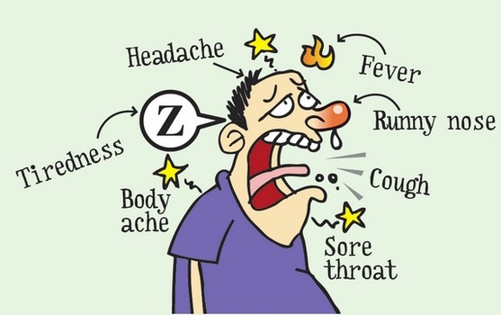
Influenza, or more commonly
referred to as the flu, is a highly contagious virus that affects the
respiratory system. Because it is so contagious, it can affect thousands of
people annually. In fact, somewhere between 5% and 20% of the United States
population will contract the virus each year (2). However, despite its
frequency of occurrence, the flu is not always a deadly disease. Of the people
who are infected, there is an average of 200,000 hospital visits a year, and,
depending on the strain, the number of flu-related deaths can range from 3,000
to 49,000 (2). The following fact is what makes the flu so dangerous though:
the virus mutates every year. Every year doctors scramble to create a vaccine
that will protect the public from the different flu strains that hit during flu
season; they do not always make the appropriate vaccine, though, and that is
when fatalities can accumulate. This leads to a pandemic.
Discoveries from recent research in the immunology literature, aimed at a general audience.
Monday, September 30, 2013
T-cells Found To Be Linked To Influenza Immunity – Could A Universal Vaccine Be On The Way?
Correlation between HLA alleles and pH1N1 mortality rates
Many people
have had the flu at last once in their life and see their doctors every year
for a flu vaccination. The vaccine is
typically trivalent, which means that it protects against three different
strains of the influenza virus: H1N1, H3N2, and an influenza B strain. This vaccine is given as an intradermal shot
meaning it is injected into the skin instead of the muscle2. Seeing as how flu season is fast approaching
I though it fitting to bring to light an article relating the flu, or more
specifically the pandemic influenza A virus strain (pH1N1) of the 2009-2010 flu
season. However, before delving into the
article I would like to provide the basic, necessary background information.
When an
antigen is engulfed by an antigen presenting cell (APC), its proteins are
broken down and a small peptide chain of about 10 base pairs is expressed so
that activated cytotoxic T-cells can destroy the infected cell3. The proteins that express these peptides are
called MHC (major histocompatabiltiy complex) class I and MHC class II proteins
which are encoded by the human leukocyte antigen (HLA) gene3. For MHC class I gene on HLA there are three
regions: HLA-A, HLA-B, and HLA-C for which many different alleles exist3. Some MHC alleles display the peptides for
T-cell recognition better than others which is why different people have
different immune responses to the same pathogen.
Additionally, CD8+ is the specific receptor that MHC class I
binds to on T-cells3.
Infected cells also release interferons (INFs) that increases the
expression of MHC molecules which helps to fight off infection by causing more
interactions with T-cells3.
There May Be Hope for a Universal Flu Shot
A recent study suggests that the development of vaccines that protect against multiple strains of Influenza A Virus (IAV) on a long-term scale is possible.1
If you
have ever stayed home from school or work with a sudden fever, cough, sore
throat, and generally achiness, you probably had the flu. You may have even
gotten the flu shot, but influenza still got the best of you. Vaccination for
the flu is difficult, because the virus mutates and develops different strains
all the time.2 The flu shot or nasal mist that we use to prevent
influenza is typically trivalent, which means it has the ability to protect
against three specific flu strains.3 These strains change depending
on what scientists predict will be the most dangerous strains to the public
that year.3 Obviously, an ideal vaccine would protect against any
form of the flu. For this reason, research that shows how CD8 T cells can be
cross-protective against heterosubtypic infections is especially promising.
The goal
of the study was to better understand the effects of vaccination on the way the
immune system responds to infection with IAV. Influenza A virus is found in
both animals and humans. It is also generally responsible for seasonal flu
epidemics in humans.4 The most at risk individuals are children, the
elderly, and other people with weakened immune systems. Influenza is
responsible for between three thousand and forty-nine thousand deaths per year,
as well as up to two hundred thousand hospital visits.4
results of the experiment, one must have a basic
understanding of certain components of the immune system, as well as specific
terms and phrases that are important in the study.
CD8
T cells are cytotoxic cells that specifically terminate the acute influenza
infection and contribute to long-term memory of the virus.1 Virus-like
particles (VLP) are developed from influenza virus proteins, but they do not
contain IAV genomic material.1 This means that resemble the actual
virus, but they are replication deficient. Because of this characteristic, VLPs
have a greater potential for use in the vaccination of high influenza risk
individuals.1 Lung-draining lymph nodes (dLNs) are the part of the
body in which naïve CD8 T cells are primed. The cells then leave the dLNs when
they migrate to the lungs (the site of infection with IAV).
The
study conducted by Hemann, Kang, and Legge made many comparisons between
control mice and mice that were vaccinated with VLPs prior to IAV infection.
The experiment was broken up into several different parts in order to show how
different aspects of immunity contributed to their overall determination.
The Power of the MHC
HLA MHC Complex onHuman Chromosome 6 |
Infectious and autoimmune diseases have been correlated to polymorphisms within the MHC. Polymorphisms are defined as different alleles of a gene existing in a population. The high levels of polymorphisms in the MHC are suggested to be due to co-evolution between host genotypes and pathogens. In other words, the “pathogen bad guys” evolve to avoid detection by the “good guys” in our bodies. The “good guys” respond though a process by which MHC genes encode cell surface proteins which bind and present peptide antigens (in the form of peptide MHC complexes) to T cells. A T cell mediated immune response (“good guys” spring into action) is then triggered if the antigen is recognized as foreign. Specifically, pathogen adaptation and virulence evolution seem to be locked in a battle with polymorphic antigen-presenting MHC genes for control of the health of the human body. It is primarily the interaction effect between the virus genotype and the host genotype that dictates variation in viral fitness and virulence, not the individual effects of virus genotype or host genotype (Table 1). This leads to specialization between a particular pathogen and host genotype.
HIV and Neurocognitive Dysfunction
HIV,
human immunodeficiency virus, infection is one of the largest public health
problems we face today. The WHO estimates that currently over 35 million people
worldwide are infected with HIV. HIV primarily infects and kills helper T
cells, cells that have the critical role of activating and supporting other cells
of the immune system, thus impairing the immune response on a broad level. It
is well known for causing AIDS, the acquired immunodeficiency syndrome, which
severely weakens the body’s immune response and leaves it open to attack by
foreign antigens. However, less widely known, HIV also causes neurocognitive
impairment and encephalitis (swelling of the brain), estimated by Heaton et al
to affect one third of individuals infected with HIV. This facet of HIV
infection, which affects a whopping 10 million people worldwide, is an important
concern and is now receiving more attention.
Recently,
the Section of Infectious Diseases of the Nervous System at NIH, led by Dr.
Avindra Nath, and their colleagues at Johns Hopkins School of Medicine have discovered
possible mechanisms for HIV-mediated neurocognitive impairment. In a recent
paper in the Journal of Neurovirology, these researchers looked at the
impairment of adult neurogenesis and neurite outgrowth in the hippocampus using
a HIV mouse model. They believe that the growth patterns of neurons in this
part of the brain, which deals with processes such as memory consolidation, may
be disrupted during HIV infection and underlie the cognitive dysfunction seen
in HIV infected patients. While previous research had found that HIV infects
glial cells and not neurons, it was shown that HIV infection lead to synaptic
pruning and apoptosis (Ellis et al 2007).
Friday, September 20, 2013
Do T Cells Have A Need for Speed?
A recent study focuses on the time
required for T cells to identify, interact with, and mount a response against
pathogens within the human body.
The life of
an infectious pathogen is not an easy one. Should a virus, bacterium, prion, or
fungus manage to make it past the body’s first line of defense – obstacles including
anatomical barriers such as the skin and physiological barriers like the hostile
low pH environment of the stomach – it still must face a barrage of innate and
adaptive immune responses before successfully establishing itself. Once a pathogen has entered the body, the innate
immune system – defined as the cells and mechanisms that defend the host
against foreign organisms in a non-specific manner - may recognize the non-self
intruder and induce an immune response. This eventually leads to the activation
of the adaptive immune system and, in turn, of white blood cells like T and B
lymphocytes (or T and B cells) which quickly identify and eliminate the
pathogen. It is the accuracy and speed of these cells’ responses that makes the
adaptive immune system so effective in protecting the body against infection. So the short answer is, yes, T cells do have a need for speed. To
contest to this, a recent study now suggests that that once a T cell senses an
antigen – a task that can take only a matter of seconds – it can decide the
fate of the invading microorganism within minutes.
This new
study, released in The Journal of
Immunology just a few months ago, tested T cells under various conditions
to judge their aptitude for identifying invading pathogens. According to the
authors, there are four factors that limit the ability of T cell to recognize
pathogens. These include:
- T lymphocyte bears a single T cell receptor (TCR). This means that any given T cell can only recognize a single antigen structure. Therefore T cells are very sensitive to interactions but are vastly outnumbered by antigen presenting cells (APCs)!
- Thousands of proteins can be used to generate the peptide - major histocompatibility complex (pMHC) on the invading cell surface. The T cell must be able to quickly recognize one to a few of these intricate complexes on the cell to identify it.
- Exposed cell pMHCs may differ by a few amino acids. Despite this, the TCR must still recognize them and be able to properly bind to trigger a T cell response.
- The TCR and pMHC complex is only ~ 14 nanometers in length. One nanometeris one-billionth of a meter, so these are miniscule complexes that must somehow closely interact in a stable way. This interaction is strongly dependent on the tiny movements made by the T cell membrane.
These limiting factors require that we
know two important things about T cells if we want to understand the speed and
specificity of the antigen detection process. Firstly, we need to know the how
often a T cell and an antigen presenting cell contact each other and for how
long. Secondly, we need to know if and how T cell membrane motions are altered
as a result of antigen detection.
Thursday, September 19, 2013
The Road to Unconventional Immune System Memory

![]() |
General Memory B Cell Development |
A
recent paper published in The Journal of
Immunology explores the road to a memory response. The two big cell types involved in the
pathway they explore are T and B cells.
The T cells involved help to activate B cells, which then produce
antibodies. These are small
proteins that circulate through the body are responsible for “tagging” foreign
bodies, or antigens, for destruction by other immune cells or neutralizing the
effect of the foreign body. Antibodies
come in different structures with different functions, and can switch
structures during the development and maturation of the B cell from a naïve
cell to memory cell. The two
structures the researchers look at are IgM, which are usually produced by naïve
cells that haven’t switched antibody type yet, and IgG, which are usually
produced by B cells that have fully matured into memory B cells. However, recent studies have shown that
there are also memory IgM B cells, but their characteristics, purpose, and
development are unclear. Immune
cells are often identified and characterized by the molecules expressed on
their cell surface. Called CD
markers, or cluster of differentiation, they allow scientists to give a unique
expression pattern to help identify and isolate new types of immune cells. In the quest to understand the memory
IgM B cells, one of the things the authors tried to undercover was a unique CD
expression pattern on these cells.
They also looked at a mouse model of human ehrlichiosis, a bacterial infection
from ticks, to explore the necessity of a T cell-B cell interaction for the
activation of these IgM memory cells.
The also used this model to look at a secondary exposure to an antigen
and determine the connection between these new memory cells and typical IgG
memory
Monday, September 16, 2013
Tiny Pests Causing Huge Problems
Why do people put on bug spray when they
go outside? The obvious answer is that having itchy mosquito bites all
over your arms and legs is one of the most irritating feelings ever.
However, most people forget that these tiny insects can carry extremely
dangerous viruses that can cause severe illness or even death. West Nile
virus (WNV), a well-known mosquito-borne virus, is the major cause of
encephalitis (inflammation of the brain), which is a disease characterized by
flu-like symptoms, and in severe cases, seizures and sensory/movement
issues. Dengue virus (DENV) is another dangerous mosquito-borne virus
that is a major cause of viscerotropic disease, which is a disease that is
associated with yellow fever and ultimately leads to organ failure and
death. These viruses cause significant problems all over the world, and
currently there are no approved vaccines to prevent or combat WNV and
DENV. In order to develop effective vaccines, we need to understand how
our bodies’ immune system recognizes the viruses and provide protection.
“Innate Immune Sensing of Flaviviruses” is a recently published paper in PLoS
Pathogens, in which Suthar et al. (2013) discusses recent research findings
on the way that the innate immune system interacts with WNV and DENV to trigger
an antiviral response.1 Also, understanding how these viruses
can evade our immune response is equally important for developing new treatments. A group mentioned in “Innate Immune Sensing
of Flaviviruses,” Aguirre et al. (2012)2, investigate one of the
main mechanisms that DENV uses to inhibit inducing an immune response.
WNV and DENV are part of the flavivirus
family, and they contain a single stranded RNA genome. In order to
recognize these viruses, we encode receptor proteins called pattern recognition
receptors (PRRs), which recognize and bind to structures on pathogens that are
not present in normal, human cells. These structures are called
pathogen-associated molecular patterns (PAMPs). Once the virus is
recognized, antiviral immune defenses are initiated which leads to
inflammation.
The two PRRs that recognize the viral,
non-self RNA structures are called retinoic-acid inducible gene-1 (RIG-1)-like
receptors (RLRs) and myeloma differentiation factor 5 (MDA5). RIG-1
recognizes and binds to double stranded RNA with a triphosphate group on the 5’
end. MDA5 interacts with long double stranded RNA. Double stranded
RNA does not exist in normal, host cells, and it is a common viral PAMP.
Once the virus is recognized, RIG-1 and MDA5 induce the production of type 1
interferon (IFN) and proinflammatory cytokines that will migrate to the site of
infection and recruit/help produce other factors needed to eradicate the
virus. A study done by Fredericksen et al. (2008) demonstrated that RIG-1
and MDA5 function cooperatively in creating an antiviral response to WNV.3
They found that RIG-1 is activated first by WNV, and then MDA5 is activated to
sustain type 1 IFN and ISG expression (initiated by RIG-1).3
It is known that the PRRs RIG-1 and MDA5 are involved in pathogen recognition
and initiate the immune response, but it is still unclear what the exact PAMP
RNA substrate and nucleic acid sequence that RIG-1 and MDA5 interacts
with.
Subscribe to:
Posts (Atom)