Many types of cancers exist today
in this world. One of the most common types of cancer found in humans is
lymphoma. Lymphoma is a type of cancer where the white blood cells called the
lymphocytes divide uncontrollably. (3) This form of uncontrollable division
leads to what are called tumors. Tumors as most of us know are masses of
abnormal cells due to an abnormal growth or division of cells. The tumor cells are your Villains because they are trying to kill you. In the event of tumor
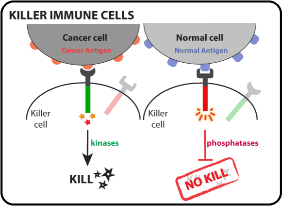
cells attacking, the immune system recruits what are called T cells. The T
cells, specifically Cytotoxic T Lymphocytes (CTLs), are the Heroes because they are trying to save
you. In a new study, researchers examined the role of CTLs in a type of
lymphoma called non-Hodgkin lymphoma, which is more common than the Hodgkin
lymphoma. Although knowing the differences is not important in being able to
understand the topic of this recent study, the differences between the two
lymphomas can be found in the reference of lymphoma. (3) The importance of this
paper lies within the interaction of the Cytotoxic T Lymphocytes and the
lymphoma tumor cells. The new study recently published in The Journal of Immunology reveals how non-Hodgkin lymphoma tumor
cells can actually “stop” the T cells. When we thought things couldn’t get any
worse when it comes to cancer. When I mean by “stop”, the amount of tumor cells
present determines a threshold at which the CTLs inactivate as well as delete
rapidly in an antigen specific manner. Within the study, the researchers try to
figure out the causes of how and what
inactivates the CTLs.
Before we begin with the paper, here
are some quick basics of immunology as well as what the research paper will be
about to get a better understanding:
CTLs are called Cytotoxic T
Lymphocytes, or CD8 T cells. They are called CD8 because they carry a surface
marker protein called CD8 that allows them to recognize a surface protein
called MHC class I (a protein expressed by all nucleated cells that presents
broadly-specific foreign peptides). For understanding purposes of the T cells,
I will be referring the T cells as CTLs just as the researchers call them in
the paper. Like I said before, CTLs are a type of white blood cell found in the
immune system. They kill cancerous cells, host cells that are infected by
viruses, or cells that are damaged in any way. The purpose of having CTLs in
your body is so that they monitor your body to destroy the person’s
infected/irregular cells by direct cell contact before infected cells can
spread throughout your body.
The CTLs kill the tumor cells in a
process called apoptosis (same process for killing infected cells) where the
tumor cells commit suicide after binding their antigens found on the surface to
the TCRs (T Cell Receptors). Depending on the structure of the proteins in the
binding sites found between the receptor and antigen, determines the
specificity of the interaction. The interaction between an antigen and a
receptor is like the binding of a lock and key.
Here is some background info on
what is involved in the paper. Reading this would be extremely helpful in
understanding what is going on:
The scientists start with two
transgenic mice to model the human non-Hodgkin lymphoma tumor cell groups with
the first being Eμ-myc-GFP, and the second being Eμ-myc-OVA.
Allow me to clarify the meaning behind
the names for the two groups. Firstly, the “Myc” is a gene being used to
research the study. The purpose of the Myc in our body is so that the Myc gene
codes for a protein that acts as a transcription factor that binds to regions
of DNA to regulate expression of other genes. When Myc is mutated, it causes
overexpression of other genes such that it ultimately leads to cancer. How
these scientists are able to manipulate the gene in order to mutate the Myc is
by having an enhancer called the IgH enhancer. Myc is a proto oncogene. (4) The
IgH enhancer, represented as the “Eμ”, acts on the Myc to develop into lymphoma
cancer cells so that the researchers can study the causes of lymphoma tumor
cells and CTL inactivation inside transgenic mice.
“OVA” is the abbreviated form of
ovalbumin. Ovalbumin is the main protein found in egg white and it is commonly
used as an antigen in immunology to stimulate an immune response to test
whether a specific immune cell will elicit a response compared to a control.
The control simply contains GFP
along with the tumor cells. GFP For those who do not know what GFP stands for
is Green Fluorescent Protein. GFP
is used frequently in biology labs in order to see if that protein will
actually give a color as an indicator of its presence.
The Eμ-myc-GFP contains tumor cells with GFP,
but no OVA mouse strain. This is the control. The Eμ-myc-OVA also contains
tumor cells, but with the addition of both GFP and OVA to see if the OVA is a
functional antigen.
The two groups both contain tumors;
however, only the OVA cell group will elicit an immune response because only
that cell group contains an antigen that will bind to the CTLs that are
specific for OVA.
Now that you have some context, knowing
the function of the “Eu-Myc” in both groups is not as important as knowing the
“GFP” and “OVA” parts because the OVA determines the antigen binding to the CTL
in this paper. So pay attention to the CTL interaction when you are reading.
Now we are getting back to the subject
of matter. The two groups both contain tumors; however, only the OVA
cell group will elicit an immune response because only that cell group contains
an antigen that will bind to the CTLs that are specific for OVA antigen. In
this paper, the CTLS are looking to kill the tumor cells that contain
ovalbumin. Those CTLs are specified as anti-OVA OT-I CTLs because they are
specific for the ovalbumin.
When the scientists injected the
anti-OVA CTLs after 3 days of tumor development, the CTLS killed off almost all
of the tumor cells. But when the scientists injected the anti-OVA CTLs after 5
days of tumor development, the tumor cells did not die off. In fact, the OVA
tumor cells in the 5-day count were about the same high numbers as the control
group in days 3 and 5. In the 3 days and
5 days of the GFP control, the tumors stayed relatively the same with high
numbers of small increases after each day due to cell division as expected
because there was no CTL specific for the control. (Fig 1)
After experimenting the
interactions between the CTLs and the tumor cells, the scientists recovered the
amounts of the CTLs in each group for both days from the spleen where all the
action took place. In order to see any type of difference in the CTL counts for
the 3 and 5-day counts for each group, lymphocytes were identified and measured
via a process called flow cytometry. Upon recovering the CTLs, only a few CTLs
were recovered from the Day 5 of the GFP-OVA mouse, but the CTLs in the GFP
alone mouse were still the same high population number compared with the day 3
results, and compared to the no-CTL injection results. (Fig 2)
The results were promising, but the
scientists needed to dive in deeper into how the CTLs were being deleted and
inactivated.
From
recovering the CTLs, the scientists also noticed the characteristics of
exhaustive T Cells and increased PD-1 receptors. Exhausted CTLs are as stated “a
process that involves the progressive loss of function and ultimately death of
the virus-specific T cells”. (2) The other characteristic that scientists
noticed were that the day 5 CTLs showed higher numbers of having PD-1
(Programmed Cell Death 1) receptor and down regulation of TCR expression. The
scientists realized then that the cause of the up and down regulation of the
receptors had to be related to antigen specific.
They knew the CTLs were antigen
specific because of the OVA. In order to confirm if this antigen specific was caused
by direct cell contact or indirect contact by cytokines, they also injected a CTL
that had characteristics of being a nonspecific antigen such as glycoprotein.
The CTL is named gBT-I CTL, which also expresses receptors that could be affected by
the inactivation as well as the OVA CTLs only if the mechanism is by the
release of cytokines. After the scientists co-injected the two CTLs, they found
that only the OVA CTL was affected. This meant the inactivation of CTLs is caused
by direct contact of the antigen because the gBTI-CTLs were unaffected. The
scientists stated that the direct contact also meant the antigen is a cognate
antigen that requires an APC (antigen presenting cell that presents the
specific antigen to the CTL).
Now that the researchers have figured
out precisely how the inactivity of
CTLs occurs as that being direct antigen specific, the question becomes what causes the inactivity. Many of the
researchers thought it could be the likes of an APC.
After figuring out the
dendritic cells were not the cause of the inactivation when they put dendritic
cells (a cell type of an APC) that were deficient in presentation with CTLs into
tumor bearing mice, they next assessed whether the tumor cells were the APCs
that inactivated the CTLs. The scientists injected a new lymphoma called bm1.Eμ-Myc-OVA
that is deficient in OVA presentation meaning that the CTL should live if the
tumor is unable to make direct contact with the CTL. When the scientists
gathered the results for the tumor cells, the CTLs were a smaller number in the
original OVA while the CTLs in the bm1 deficient presentation of OVA were at
the same amount as those CTLs in the GFP control meaning the CTLs lived. This
showed that the CTLS were active, but the tumor cells still had the same population,
as those in the GFP control because the CTLs were unable to make contact to try
to kill the tumors since the tumors were deficient in peptide
presentation.
Afterwards,
the scientists then tried to see if the tumor cells resisted the chemicals the
CTLs release when they engage the tumor cell. The chemicals the CTLs release
are called interferon- γ, and kill the tumor cells. To see if the tumor cells acquired a
resistance after the contact inactivation of CTLs, they injected more CTLs a
day after the 5 -day count at a smaller tumor population. As a result, the
tumors ended up dying meaning that the tumors did not acquire resistance to
CTLs after the initial contact for inactivation of CTLs. Since tumor cells do
not create a resistance, then the tumor cells can inactivate the CTLs at a
certain threshold.
Now the scientists test to see what
affect the chemotherapy drug CTX, also called cyclophosphamide, has on the
tumor cells with CTLs. The scientists wanted to compare the affects of a low
dose and high dose of the chemotherapy on the tumor cells to see if the CTLs
inactivate depending on the dosage level. When the scientists injected CTLs
after a low dose, the tumor cells quickly bounced back to eventually inactivate
the CTLs. But, when the scientists placed CTLs after a high dosage of
chemotherapy on a high population of tumor cells, the CTLs were effective in
eliminating the rest of the lymphoma tumor cells. This showed the CTLs become
inactive at a certain threshold, but at the same time show another treatment
that is successful in eliminating tumor cells. Now that they identified the
problem for the most part………what is next?
Even though these scientists did not
find the exact answer they were looking for in terms of the exact mechanism for
inactivation, they did find a more efficient treatment for patients with
lymphoma, and that is by supplying CTLs directly after the chemotherapy so that
the large tumor counts are reduced enough to a point where they will no longer
be able to reach the threshold to inactivate the CTLs.
The researchers noted that uninfected
host cells carry mechanisms to inactivate the activity of CTLs so that the CTLs
will not damage any of the individual’s vital tissues (as a tolerance
mechanism). Given that, the researchers believe the CTLs are being inactivated
due to an autoimmune prevention mechanism. As a result of this mechanism, it
appears that the mechanism is still preserved within the tumor cells since the
tumor cells used to be normal host cells before they became tumor cells. As a
result of that hypothesis, it would be interesting to further the research by
comparing those CTLs that recognizes viruses/tumors being compared to those
anti-self CTLs to see if the anti-self CTLs will inactivate as an autoimmune prevention
when it is in the presence of tumor cells.
Primary Source:
1)Prato, Sandro et. Al. Rapid Deletion and Inactivation of CTLs upon
Recognition
of a Number of Target Cells over a Critical Threshold. 9 September
2013. Journal Immunology, 191, 3534-3544.
Online Publication.
Link:
Secondary Sources:
T Cell exhaustion:
c-Myc:
Lymphoma:
Images:
No comments:
Post a Comment