-->

Monday, December 2, 2019

Looking for Antibodies: New Development in Uncovering the Cause of Pediatric Acute Flaccid Myelitis


In response to: Schubert, R. D., Hawes, I. A., Ramachandran, P. S., Ramesh, A., Crawford, E. D., Pak, J. E., ... & Lyden, A. (2019). Pan-viral serology implicates enteroviruses in acute flaccid myelitis. Nature medicine, 1-5.

Over the past seven years, a polio-like disease called pediatric acute flaccid myelitis (AFM) has plagued children in the United States in increasing numbers. The first case of AFM was reported in California in 2012, but since then, there have been over 500 reported cases across the United States (Ayscue et al., 2014). The symptoms of AFM include weakness in the arms and legs, difficulty breathing, and internal lesions to the child’s spinal cord (Sejvar et al., 2016). While many children who develop AFM can survive the infection, very few children are able to make a full recovery; as a result, most afflicted children must live with permanent limb and respiratory disabilities (Sejvar et al., 2016). Currently, medical researchers and neurological pediatricians do not know what causes the disease, so understanding why this debilitating disease manifests is of critical importance to the medical community.
While the cause of the disease is unknown at this time, epidemiological research, which looks at broad trends in public health and diseases to make connections in regards health-related questions, has linked AFM to two strains of enterovirus, EV-D68 and EV-A71. Enteroviruses are single-stranded positive-sense RNA viruses and are a genus of virus from the Picornaviridae virus family. Strains of enterovirus are the causative agents for a variety of diseases ranging from the common cold to the poliovirus. The EV-A71 strain of the enterovirus has been associated with a spectrum of neurological diseases while the EV-D68 strain has been primarily found in children suffering from respiratory illnesses such as pneumonia or bronchiolitis (Brown et al., 2018). In 2014, spikes in AFM cases in the United States were found in areas that also had increased rates of EV-D68 and EV-A71 infection (Aliabadi et al., 2016). While the epidemiological research has demonstrated an association between both AFM and EV-D68 and EV-A71, no studies have been able to produce results that would indicate a causal relationship between either strain of the enterovirus and AFM. To determine a causal relationship between a virus and a disease, researchers generally look for the presence of either viral nucleic acid or viral antibodies in the patients’ cerebral spinal fluid (CSF). The presence of either molecule in a person’s CSF indicates that the person’s immune system is either actively fighting the virus or has previously encountered that virus. Previous research, however, has found that only two percent of AFM patients have detectable levels of enterovirus nucleic acid in their CSF. These findings have caused some scientists in the medical community to question whether enterovirus infection is the cause of AFM (Messacar et al., 2016). To demonstrate whether or not the enterovirus is the main trigger for AFM, researchers at the University of California, San Francisco performed a large scale search for viral antibodies in the CSF of patients with AFM to determine whether there is any biological evidence of a relationship between enteroviruses and AFM.
To discover which type of viruses could be linked to the development of AFM, the researchers examined the CFS of forty-two patients with AFM and fifty-eight healthy controls. Researchers screened for viral antibodies and viral nucleic acids in the CSF of both subject groups using several bioinformatics programs. These programs can be used to identify molecules in the subjects’ CSF through genome sequencing. A metagenomics next-generation sequencing (mNGS) was performed on the CFS of both the controls and the AFM patients to detect the presence of viral RNA. During the mNGS, nucleotides retrieved from the CSF of the patients and the controls were sequenced to a high degree of specificity and compared to the library of previously sequenced microorganisms and viral RNA. Following the mNGS sequencing, only one of the children with AFM tested positive for EV-A71 RNA and none of the children tested positive for EV-D68 RNA. The researchers’ results were consistent with earlier studies that had failed to find significant levels of enterovirus RNA in the CSF of children with AFM (Messacar et al., 2016).
After the viral RNA sequencing failed to demonstrate any relationship between enteroviruses and AFM, the researchers directed their attention to screening for antibodies in the patients’ CSF. Following infection, humans’ immune systems produce antibodies to target the infection. An antibody is an immune protein that specifically targets individual pathogens for they have antigen-binding sites that only attach to one type of antigen peptides that is created by solely the antibody’s target. The presence of a specific viral antibody in a patient’s CSF indicates a previous infection with the virus. Thus, although the researchers were unable to find evidence linking AFM to enterovirus infection when searching for elevated levels of enterovirus RNA, if enterovirus antibodies were found in the CSF of AFM patients the virus could still potentially be the causative agent of the disease.
To detect viral antibodies, the researchers utilized VirScan sequencing which uses bacteriophages that display viral peptides on their exterior to determine whether there are elevated levels of any viral antibody in a subject’s CSF. The VirScan program has 481,966 unique peptide sequences that can be used to identify vertebrate, mosquito-borne and tick-borne viral sequences. If the bacteriophage expressing a particular viral peptide is encountered by an antibody for the virus, the antibody will bind to the bacteriophage and the computer program which analyzes that data will signal a positive hit for the virus. The VirScan results showed that when compared with the CSF of healthy controls, the CSF of children with AFM had elevated levels of Picornaviridae peptides, the virus family to which the enteroviruses belong. When the Picornavirididae peptides were further examined, the greater levels of enrichment were almost entirely due to increased levels of enterovirus antibodies (Figure 1). The researchers also found that protein sequences common to enterovirus were elevated in the AFM patients’ CSF when compared with the controls’ CSF. For example, levels of viral protein 1 (VP1), a viral capsid protein predominately expressed by enteroviruses, were elevated in the CSF of AFM patients when compared to healthy controls (Figure 2).

Figure 1. Quantification of Enterovirus Enrichment in the CSF of Healthy Controls and AFM Cases. Following VirScan sequencing, the levels of enterovirus enrichment and the number of patients with enriched levels of enterovirus were quantified using a violin plot. The black dashed line represents the mean proportion of reads of enterovirus in the CSF and the red dotted line demarcates the third and first quartile for the proportion of reads of enterovirus. The width of the plot increases when more subjects who test positive. The greater the height of the violin plot indicates a greater proportion of positive reads in the subjects. Of the 42 AFM cases, 29 (69%) of the cases tested positive for EV antibodies. Of the 58 healthy controls, 4 (7%) of the controls tested positive for EV antibodies. There was a significant difference in the number of subjects in the two groups with the enterovirus antibodies in their CSF.

Following the VirScan search, the researchers performed an enzyme-linked immunosorbent assay (ELISA) to further assess whether enterovirus antibodies were upregulated in AFM patients. In an ELISA, antigens of interest are stuck to the bottom of plastic laboratory wells and then fluid containing a possible antibody, which for these experiments was the CSF of the AFM patients and the controls, is placed in the wells. If an antibody that binds to the antigen is present in the fluid added to the wells, the antibody will stick to the antigen and will not be removed from the plate when the nonspecific antibodies are washed away. Fluorescent substrates are then added to the wells and the amount of fluorescence in each well is used to quantify the level of antibody present in the cells. Greater levels of fluorescence in the wells indicate there were higher levels of the well antigen’s antibody present in the fluid added to the well. The researchers at the University of California, San Francisco used the VP1 protein as their well antigen and ran the ELISA with the CSF from the AFM patients and the healthy controls. Again, the researchers found that the CSF of AFM patients had elevated levels of enterovirus antibodies when compared with healthy controls. Also, AFM patients who had previously tested negative for enterovirus antibodies in the VirScan sequencing tested positive for enterovirus antibodies in the ELISA. This discrepancy in identification between the two assay calls into question the validity of VirScan for detecting enterovirus antibodies, suggesting that the researchers need to evaluate whether or not VirScan is an appropriate technique for enterovirus infection. In both the VirScan sequencing and ELISA, researchers did not find that one strain of enterovirus was predominately expressed in the CSF of patients with AFM. Researchers found that the CSF of AFM patients tested positive for multiple strains of enterovirus which indicates that co-infection with multiple strains of enterovirus might be necessary for the development of AFM, or a person who develops AFM may be more susceptible to additional infection of enteroviruses.

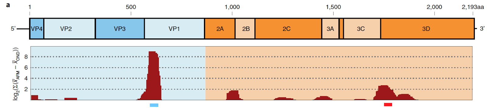
Figure 2. Enterovirus Enrichment at Specific Genome Sites in EV-A71 in the CSF of Healthy Controls and AFM Cases. Following VirScan sequencing, enterovirus-derived peptides found to be enriched in the CSF of AFM patients were plotted on genome sequence for different strains of enterovirus. The figure above demonstrates the enrichment of specific peptides found in the EV-A71 strain of the enterovirus. The BLASTp computer program was used to match viral peptides with their appropriate site in the EV-71A genome. The larger red peaks display greater enrichment of viral peptides in AFM patients when compared with healthy controls. Peptides such as VP1 were found to be significantly enriched in AFM patient’s CSF when compared to controls’ CSF.

Ultimately, the work completed by researchers at the University of California, San Francisco has provided another crucial link in establishing the relationship between enterovirus and AFM. The presence of elevated levels of enterovirus antibody in the CSF of AFM-afflicted children provides strong evidence to support that a causal relationship exists between enterovirus infection and the development of AFM. Moreover, the research shows that the detection of enterovirus antibodies in suspected patients’ CSF can be used as a diagnostic tool for identifying cases of AFM. While the work of the researchers at the University of California, San Francisco provides support for the theory that enterovirus causes AFM, more research needs to be completed before a causal link between enteroviruses and AFM is established. First, the study design of the researchers needs to be improved. The controls used for this study were significantly older than the AFM patients. The average age of the AFM patients was 38 months whereas the average age for the controls was 120 months. The age difference between the two groups presents a potential confounding variable that may have misconstrued the study results. Additionally, cohort studies, where healthy children are followed and evaluated to see which children become infected with the enterovirus, AFM, or both enterovirus and AFM, need to be completed to further determine whether or not a causal relationship exists between the two viruses for such studies allow researchers to establish a temporal relationship between the infection and the development of the disease. The proposed study would cost a significant amount of money so greater resources from agencies such as the Center for Disease Control would need to be directed towards AFM research.

AFM researchers also need to explain why enterovirus RNA cannot be detected in the CSF of AFM patients. Failure to account for why AFM patients do not present with elevated levels of enterovirus RNA in their CSF creates a major obstacle for demonstrating a causal relationship between enterovirus infection and the development of AFM. Lastly, AFM researchers should focus on uncovering a mechanism that explains how enterovirus infections result in the development of the neuro-motor deficits seen in patients with AFM. Overall, a significant amount of research needs to be completed to definitively link enterovirus infection to AFM, but the recent work completed at the University of California, San Francisco indicates that significant advances are being made in the medical community to identify the cause of this debilitating neurological disease. For further reading on the human cost of AFM infection, Pam Belluck, a reporter for the New York Times, recently detailed the experience of families with children who have developed AFM in this compelling article.

References:

Aliabadi, N., Messacar, K., Pastula, D. M., Robinson, C. C., Leshem, E., Sejvar, J. J., … Dominguez, S. R. (2016). Enterovirus D68 Infection in Children with Acute Flaccid Myelitis, Colorado, USA, 2014. Emerging infectious diseases, 22(8), 1387–1394. doi:10.3201/eid2208.151949

Ayscue, P., Van Haren, K., Sheriff, H., Waubant, E., Waldron, P., Yagi, S., … Centers for Disease Control and Prevention (CDC) (2014). Acute flaccid paralysis with anterior myelitis - California, June 2012-June 2014. MMWR. Morbidity and mortality weekly report, 63(40), 903–906.

Brown, D. M., Hixon, A. M., Oldfield, L. M., Zhang, Y., Novotny, M., Wang, W., … Scheuermann, R. H. (2018). Contemporary Circulating Enterovirus D68 Strains Have Acquired the Capacity for Viral Entry and Replication in Human Neuronal Cells. mBio, 9(5), e01954-18. doi:10.1128/mBio.01954-18

Messacar, K., Schreiner, T. L., Van Haren, K., Yang, M., Glaser, C. A., Tyler, K. L., & Dominguez, S. R. (2016). Acute flaccid myelitis: A clinical review of US cases 2012- 2015. Annals of neurology, 80(3), 326–338. doi:10.1002/ana.24730

Sejvar, J. J., Lopez, A. S., Cortese, M. M., Leshem, E., Pastula, D. M., Miller, L., … Feikin, D. (2016). Acute Flaccid Myelitis in the United States, August-December 2014: Results of Nationwide Surveillance. Clinical infectious diseases : an official publication of the Infectious Diseases Society of America, 63(6), 737–745. doi:10.1093/cid/ciw372

Sunday, December 1, 2019

Inhibiting HIV-1 Infection by Shooting the (CCR5) Messenger

Paper: Boncompain, G., Herit, F., Tessier, S., Lescure, A., Del Nery, E., Gestraud, P., Staropoli, I., Fukata, Y., Fukata, M., Brelot, A., Niedergan, F., and Perez, F. (2019). Targeting CCR5 trafficking to inhibit HIV-1 infection. Science Advances, Vol. 5, No. 10, eaax0821, doi: 10.1126/sciadv.aax0821

       Plasma membrane receptors are a common feature found on cell surfaces across the body. These proteins, which are typically embedded directly in the cell membrane, can serve many functions, including cell-to-cell communication and identification to the immune system. Unfortunately, their exposure to the extracellular environment makes them a frequent target for infectious pathogens. By binding to receptors found on the surface of specific cell lines, viruses and other microorganisms can identify and target certain cells for infection and propagation.
  Two of these membrane receptors in human immune cells, CD4 and CCR5, are targeted and bound by HIV-1, allowing the virus to properly identify and enter white blood cells. A small portion of the population, however, has a rare genetic mutation that results in CCR5’s absence at the cell surface, leading to HIV-1 infection resistance (1). Because of this, scientists have begun to develop anti-HIV therapies that target CCR5 expression. The most successful of these therapies binds directly to the CCR5 receptor, preventing HIV-1 binding and infection (2). This discovery has led researchers to investigate how the disruption of the transport of the CCR5 receptor to the cell membrane may provide an alternative method of HIV-1 treatment.
  Before a membrane receptor is present in a cell surface, it must be synthesized and transported in a series of cell organelles that comprise its secretory pathway. This includes the Endoplasmic Reticulum (ER), where proteins are produced, the Golgi apparatus, which packages and ships proteins, and vesicles, which enclose the proteins and fuse to the cell membrane. By disrupting this pathway for CCR5 receptor delivery, scientists in a recent study were able to prevent its presence on the cell surface and therefore stop virus binding and HIV-1 infection.
  To first gain an understanding of the secretory pathway of CCR5, the researchers, which represented the Curie Institute and the University of Paris, compared the receptor's membrane transport to that of tumor necrosis factor (TNF), another cell receptor. To accomplish this, they used an assay called RUSH which allows for the synchronized release of proteins in the secretory pathway after the addition of a molecule called biotin (3). Using this method, the researchers could time the transport of the proteins from one organelle to another. To visualize the movement of the proteins, they used a method called immunofluorescence, which labels the protein of interest with a dye that fluoresces under a light microscope. Together, these methods allowed them to observe and contrast the secretory pathways of the CCR5 and TNF protein receptors.
  Using HeLa cells expressing CCR5 and TNF, the researchers induced transport with biotin and observed very different transport kinetics between the receptors. Visualization of the receptors in cells at 0, 15, and 20 minutes showed that CCR5 reached the plasma membrane more slowly and through different intermediate structures compared to TNF (Figure 1B,C). To confirm this kinetic transport difference, they used flow cytometry to measure the surface fluorescence intensity over time. This measurement found that CCR5, while transported more slowly than TNF, is more stable at the cell surface (Figure 1D). Graphically, this is seen by TNF intensity rising more rapidly than CCR5 but then almost immediately dissipating. These results indicate that CCR5 and TNF have different transport characteristics that are likely maintained by distinct molecular machineries that can be selectively targeted.

Figure 1. Transport Kinetics of CCR5 and TNF Receptors. (A) Immunofluorescence of HeLa cell expressing both CCR5 and TNF receptors (Top), CCR5 only (Middle), and TNF only (Bottom) at 0, 15, and 20 minutes after biotin addition. (C) Magnification (x2.8) of Golgi complex region. (D) Kinetics of arrival of CCR5 (magenta) or TNF (cyan) to the cell surface after release from the ER measured by fluorescence intensity.

       To search for molecules that may be candidates for inhibiting CCR5 secretion, HeLa cells expressing CCR5 and TNF were incubated with thousands of drugs from two chemical libraries: 1200 drugs from Prestwick Chemicals and 2824 drugs from the U.S. National Cancer Institute (NCI). By observing the transport of CCR5 and TNF by immunofluorescence, they determined which molecules inhibited transport for either or both receptor proteins. This allowed researchers to identify a small subset of fifteen molecules that specifically inhibited CCR5 transport.
  Once a list of molecules that inhibited CCR5 was obtained, the researchers performed experiments on the specificity of the molecules, observing if any inhibited transport of the closely related CCR1 and CXCR4 receptors. If the molecules also inhibited these closely related receptors, they could be ruled out as potential CCR5-specific therapeutic candidates.
  To observe the effect of the molecules on the transport of the closely related CCR1 and CXCR4 receptors relative to CCR5, they again used immunofluorescence to label the receptor proteins. This allowed surface fluorescence to be measured so they could determine whether receptor transport was inhibited in the cells. These surface expression results were graphed relative to two controls. The first control consisted of just a dimethyl surfoxide (DMSO) solution, which contained just the dissolved molecules and allowed transport to occur uninhibited. The second control did not induce transport with the addition of biotin, resulting in little to no surface fluorescence. This experiment yielded three potential molecules that strongly inhibited CCR5 (Figure 2B) while also minimally inhibiting CCR1 and CXCR4 (Figure 2C,D). In these graphs, surface expression closer to 0 indicated that the receptor transport was inhibited while surface expression closer to 100 indicated little no inhibition. These three molecules were termed molecules 13, 14, and 15 throughout the paper and across the figures.

Figure 2. Molecules 13, 14, and 15 inhibit CCR5 transport and do not inhibit CCR1 or CXCR4 transport. (A) Kinetics of synchronized transport of three chemokine receptors-CCR5 (black), CCR1 (red), and CXCR4 (green)-to the cell surface using the RUSH assay. Receptor transport was induced by the addition of biotin at time 0. Surface expression fluorescence intensity measured at end-point (2 hours) of the effects of the CCR5 inhibitory molecules on the trafficking of CCR5 (B), CCR1 (C), and CXCR4 (D) in HeLa cells.

  Once three candidate molecules were found to specifically inhibit CCR5, their potential therapeutic application against HIV-1 was finally tested. To accomplish this, the researchers first confirmed that the molecules inhibited CCR5 transport in human immune cells rather than just HeLa cells. After this was demonstrated, they then tested if HIV-1 infection decreased in these cells with the addition of the molecules.
  Confirmation that the molecules inhibited CCR5 transport in immune cells was performed by again measuring surface expression of the receptor. However, the immune cells were isolated from various healthy blood donors. This experiment found a reduction in the surface expression of the receptor, confirming that CCR5 receptor transport was inhibited in living human immune cells.
  To test if any of the molecules decreased HIV-1 infection, they used an assay called BlamM-Vpr (BV) which detects viral entry into cells by a change in protein fluorescence (4). Observing HIV-1 entry in immune cells with the addition of the three molecules, the researchers found all molecules to reduce cell infection by 45.7 to 78.0% (Figure 3C). To confirm that the molecules’ inhibitory effects were due to the absence of CCR5, a CCR5-independent HIV-1 virus termed VSVG-pseudotyped virus was used a control. As expected, this virus experienced no significant difference in virus entry into the immune cells, able to enter cells without the CCR5 receptor (Figure 3D). As an additional means of measuring HIV-1 infectivity, they quantified the total amount of viral protein produced by infected immune cells. HIV-1 viral production and secretion were both strongly reduced by 31.4 to 76.0% as a result of the addition of the molecules (Figure 3E).

Figure 3. Treatment with molecules 13, 14, and 15 decrease HIV-1 infection in human immune cells. Inhibition of entry of HIV-1ADA (C) or HIV-1VSVG (VSVG pseudotyped) (D) containing BlaM-Vpr (BV) with immune cells mediated by compounds. Fraction of viral protein produced by HIV-1ADA infected cells relative to control (E). Each black point represents one donor analyzed independently.

  The results of this study have significant therapeutic implications for patients infected with the HIV-1 virus. By disrupting the presence of the CCR5 receptor at the surface of immune cells, HIV-1 infection can be inhibited. Assessing the effect of thousands of molecules on CCR5 transport and further testing their effects on other receptor transport yielded three molecules that specifically disrupted the CCR5 receptor’s secretory pathway. Immune cells treated with these molecules showed a significant reduction in HIV-1 infection (Figure 3). These findings suggest that these molecules could serve as potential drug candidates for treating HIV-1 and therefore help prevent its development into AIDS.
  This paper offers exciting new insight into using the secretory pathway of proteins as a means of treating medical problems on a cellular level. Because little is known about protein-specific regulation of transport in cells, there is huge potential for new medical treatments to arise with further research. Many of these treatments could follow the lead of this study and disrupt the secretion of receptors that viruses rely on for infection. However, the full potential of using the secretory pathway to prevent pathogenesis is yet to be discovered.

1. Liu, R., Paxton, W.A., Choe, S., Ceradini, D., Martin, S.R., Horuk, R., MacDonald, M.E., Stuhlmann, H., Koup, R.A., and Landau, N.R. (1996). Homozygous defect in HIV-1 coreceptor accounts for resistance of some multiply-exposed individuals to HIV-1 infection. Cell, 86:367–377, doi:10.1016/s0092-8674(00)80110-5

2. Tebas, P., Stein, D., Tang, W.W., Frank, I., Wang, S.Q., Lee, G., Spratt, S.K., Surosky, R.T., Giedlin, M.A., Nichol, G., Holmes, M.C., Gregory, P.D., Ando, D.G., Kalos, M., Collman, R.G., Binder-Scholl, G., Plesa, G., Hwang, W.T., Levine, B.L., and June, C.H. (2014). Gene editing of CCR5 in autologous CD4 T cells of persons infected with HIV. N. Engl. J. Med., 370:901–910, doi: 10.1056/NEJMoa1300662

3. Boncompain, G., Divoux, S., Gareil, N., de Forges, H., Lescure, A., Latreche, L., Mercanti, V., Jollivet, F., Raposo, G., and Perez, F. (2012). Synchronization ofsecretory protein traffic in populations of cells. Nat. Methods, 9:493–498, doi: 10.1038/nmeth.1928

4. Cavrois, M., De Noronha, C., Greene, W.C. (2002). A sensitive andspecific enzyme-based assay detecting HIV-1 virion fusion inprimary T lymphocytes. Nat. Biotechnol., 20:1151–1154, doi: 10.1038/nbt745

Shape-Shifting Virus Poses Vaccine Difficulty


Dengue virus is a plus-strand RNA virus that infects approximately 400 million people annually, especially in the tropics of South and Southeast Asia.1 As a plus strand RNA virus, the genome that enters the infected host cell can act directly to start translating proteins, making the replication and proliferation of more viral particles rapid as compared with many other types of viruses. Dengue poses a serious health threat to a large proportion of the world as it can cause hemorrhagic fever and shock. The human immune system attempts to eradicate the virus, however, it faces multiple complications as the virus is constantly evolving to escape the immune system.
It is extremely difficult to create a vaccine for dengue virus for three reasons. Primarily, there are 4 circulating serotypes in the human population. What this means is that not every type of dengue virus is exactly the same and, therefore, the body’s response to each serotype is slightly different, making the production of a successful vaccine difficult. Another reason why it is difficult to create a vaccine is because there is evidence that dengue can rely on antibody-dependent enhancement (ADE). ADE occurs when a slightly different version of the virus becomes more competent at infecting hosts because similar antibodies are able to bind to the virus, but not able to neutralize it, facilitating viral entry into cells.2 What’s an antibody, you ask. Antibodies are one way the human immune system recognizes specific infections based on structure and responds to block the virus from entering cells. Finally, there is more recent evidence that certain serotypes of the virus can infect individuals with 2 distinct shapes that are temperature dependent. The outer protein, or envelope protein, is what the antibodies recognize and bind to in a highly specific manner.3 When the shape of the envelope protein changes, the antibodies can no longer bind as tightly. This paper examines the reasons why there are multiple different structures for the dengue virus.
The virion, or shell of the virus, is constructed from 180 copies of the E protein encoded by the viral genome. In the “smooth” virion, the E proteins form dimers and “rafts” whereas in the “bumpy” virion, the inter-dimer and raft interactions, and some intra-dimer interactions are broken. It was found that DENV2 (dengue) will initiate the switch to the bumpy virion at either 37°C or 40°C, dependent on the strain. It was found that the strain New Guinea C-2 (NGC-2) switched their virions to become bumpy at 37°C while NGC-1 remained smooth at the same temperature. The E protein between these two strains was found to be different at 5 amino acids, or protein building blocks. The researchers created mutations at these identified amino acids to discover which one was most important at initiating the virion change from smooth to bumpy. It was found that a mutant with a single, fairly minor change, at the sixth amino acid was sufficient to confer the change from smooth to bumpy at 37°C in the NGC-1 strain without changing any other proteins. The 6th amino acid is present at the interface between dimers, suggesting the NGC-2 bumpy strain is less stable than than NGC-1.
Figure 1: NGC-1 dengue virus remains "smooth" at both 29°C and 37°C while NGC-2 changes from smooth at 29°C to "bumpy" at 37°C.  

Furthermore, the authors observed whether there were differences in the replication cycles of the different strains. They found that after 24 hours post infection, the bumpy virus had higher growth rates than the smooth virus. At 40°C, all of the virions, no matter the amino acid sequence would switch to the bumpy phenotype. This is significant to the replication cycle because dengue causes fever and as the body temperature rises, the virus is actually becoming more effective as it changes to the bumpy phenotype. In additional amino acid change was found to be important in clinical strains of the virus at position 262. This amino acid is present at the intra-dimer interface of the E protein. This demonstrates that dimerization of the E protein is necessary for the smooth conformation at 37°C. Similar to the replication rate results, bumpy virions were also better at attaching and entering into human cells.
Figure 2: Mutants 4 and 1, containing the amino acid change at position 6 and a bumpy conformation at 37°C, have the highest replication rates. Mutants that remain smooth at 37°C have lower replication rates. 

            Interestingly, previous studies have found that DENV infectivity decreases with increased temperature, contrary to the evidence that bumpy virions have greater attachment and entry and faster replication. The authors suggest that this decreased infectivity may be due to the decreased stability at increased temperatures. Another potential reason for decreased infectivity at high temperatures may be due to the greater structure of the bumpiness that antibodies would be able to bind to. An in vivo study would better be able to demonstrate how the bumpy and smooth phenotypes interact with the immune system at these increased temperatures. Future research should investigate the mutant virions in vivo to confirm whether the bumpy or smooth virus would be more successful at high, feverish temperatures. 
_________________________________________________________________________________

Lim, X., Shan, C., Marzinek, J. K., Dong, H., Ng, T. S., Ooi, J. S. G., … Lok, S. (2019). Molecular basis of dengue virus serotype 2 morphological switch from 29°C to 37°C. PLoS Pathogens, 15(9), 1-25. 

Halstead, S. B. & O’Rourke, E. J. (1977). Dengue viruses and mononuclear phagocytes: infection enhancement by non-neutralizing antibody. Journal of Experimental Medicine, 146, 201-217.

Bhatt, S., Gething, P. W., Brady, O. J., Messina, J. P., Farlow, A. W., … Hay, S. I. (2013). The global distribution and burden of dengue. Nature Letter, 496, 504-507. 

Fibriansah G, Tan J.L., Smith S.A., de Alwis A.R., Ng T.S., Kostyuchenko V.A., et al. A potent anti-dengue human antibody preferentially recognizes the conformation of E protein monomers assembled on the virus surface. EMBO Mol Med. 2014; 6(3):358–71.

Antiviral Actions of Tetherin Against Ebola Virus Microvesicles and Immunomodulation


          Ebola virus infection is associated with fatal diseases in humans. For example, Ebola hemorrhagic fever is distinguished by the pathological onset of severe inflammation and internal bleeding within infected hosts [2]. Despite a high prevalence of human-human transmission, Ebola is a “zoonotic” virus, meaning it enters the human population via an animal host. For this particular virus, the animal host is a fruit bat, as certain species have been shown to act as reservoirs— carrying an asymptomatic infection able to manifest as pathogenic when transmitted into humans [3]. Until 2014, Ebola was endemic, or regularly found within the population, in West Africa. However, when a mutant strain of the virus emerged, an epidemic was established in the region. This outbreak lasted until 2016 and during this period the virus found its way to eleven individuals within the U.S., which resulted in two fatalities of American citizens [4]. Today, the Democratic Republic of the Congo is currently facing an outbreak of its own, keeping research on the virus and potential ways to control its spread at the forefront of global public health efforts [5].
In terms of structure, the Ebola virus genome is composed of single-stranded, minus-sense RNA that must be converted to positive-sense by its own polymerase before translation can occur. Additionally, its lipid membrane envelope includes just one type of protein, known as a glycoprotein (GP), on its surface. This protein protrudes from Ebola viral particles, playing a key role in the attachment and fusion of the particles to host target cells. In its mature form, GP exists as two subunits: GP1, which includes a receptor binding domain for attaching to target cell surfaces, and GP2, which includes a fusion peptide and transmembrane domain to facilitate this attachment during infection. In the face of such viral infection, host immunity often works in opposition to the invading virus, creating an arms race as each one attempts to overcome the other. For instance, the host restriction factor, tetherin, is an antiviral gene regulated by the innate immune system, or the host’s nonspecific defense that kicks in directly following detection of a virus. Known to function in the suppression of many enveloped viruses, tetherin works to interfere with a virus’s ability to release progeny viral particles from infected cells in order to spread within the host. Specifically, as seen below, tetherin’s unique double-anchored structure inserts itself into both the viral envelope and infected cell membranes in order to anchor budding virions to the cell and thereby prevent free release into the host. 

Tetherin: Traps viral-associated vesicles, as pictured, or progeny virions at the cell surface by embedding one anchor into the releasing membrane and the other into the host cell plasma membrane. [1]

 
           In this study, Nehls et al. were mainly focused on tetherin’s targeting of vesicle release, in addition to trapping budding virions. Previous studies these researchers built upon have reported that, like many viruses, Ebola virus infection releases microvesicles upon infection as a mechanism to emit viral proteins or nucleic acids from infected cells. Specifically, Ebola utilizes these vesicles to carry out the release of its GP protein. To better elucidate the role of these “GP-virosomes” in Ebola’s ability to infect hosts, researchers analyzed these particles and hypothesized that tetherin interacts with the GP-associated vesicles, ultimately interfering with their release. 
            To specifically analyze GP-virosomes, as opposed to exosomes and smaller vesicles, researchers first used a centrifugation technique that separated the seemingly larger virosomes from other smaller vesicles thought to be lacking the GP viral protein. To verify that these virosomes were both vesicles of larger size and carriers of the Ebola virus GP, a western blot, as shown below, was performed showcasing enriched GP in microvesicles (pelleted at 21000xg) as compared to in exosomes and smaller vesicles (pelleted at 100000xg). 

Figure 1C: Verifying Ebola virosomes as GP-containing, non-exosomal vesicles. Supernatants of transfected 293T cells were spun down at different centrifugation speeds and GP levels were detected via western blot. Microvesicles (100-1000nm in size) pelleted at 21000xg and exosomes (<100nm) pelleted at 100000xg.

            After observing via electron microscopy that the release of these GP-virosomes from host cells is similar to the budding of progeny virions, researchers aimed to investigate tetherin’s impact on this release. First, a human embryonic kidney cell line (HEK-293T) commonly used for transfections, or the introduction of DNA to eukaryotic cells, was used to analyze the coexpression of tetherin and GP in cell culture. To track and quantify the amount of GP released by virosomes, researchers performed a western blot on the cell culture media, as this is where the virosomes are expected to be located after successful release from infected cells. However, if tetherin was expressed as well, as shown below, this release of Ebola virus GP-virosomes detected within the media was significantly reduced. This result was replicated in HeLa cells, as this cell line contains endogenous levels of tetherin, and therefore verifies that the suppression of release seen is not an unintended impact of tetherin overexpression in the transfected 293T cells. 
           
Figure 4a: Monitoring the release of GP-virosomes. 293T cells were transfected to express varying amounts of tetherin alone (mock) or in addition to cotransfection with GP. As tetherin expression increased, GP-virosome detection in media decreased in GP-transfected cells. 

            Next, researchers analyzed tetherin mutants to narrow down which components of the protein’s unique anchor shape are vital for its ability to suppress GP-virosome release. Tetherin mutants lacking the C-terminal anchor were discovered via western blot to be incorporated into the cell culture media. This result suggested that the C-terminal anchor facilitates tetherin insertion into the host cell plasma membrane and is therefore vital for the protein’s ability to prohibit virosome release. Without this region, tetherin is not only unable to keep GP-virosomes from entering the cell culture media, but is also incorporated into the virosomes themselves. After narrowing down the vitality of tetherin’s shape to its antiviral function, transfections with tetherin derived from various animals, from hamsters to alligators, were compared to human tetherin functioning. Differential levels of GP detected in the media based on tetherin variation supported a conclusion that the primary sequence of tetherin, as it varies by species, is also important for the efficiency of the protein’s antiviral activity. Turning their sights to regions of the Ebola virus GP important for virosome release, researchers then analyzed the impact of various mutations in either the receptor binding or transmembrane domains of the protein. After transfecting HeLa cells with a GP variant containing a change in its transmembrane domain, tetherin levels were detected via a proximity ligation assay that uses antibodies against tetherin to fluorescently image the protein, as shown in the figure below. Levels of tetherin detected in the GP variant cells (ELE) were significantly reduced as compared to cells with wild-type GP transmembrane domains. This finding implied that this region of the Ebola virus GP is essential to tetherin’s ability to interact and prevent the release of GP-virosomes. 

Figure 5c: The GP transmembrane domain is important for tetherin binding. HeLa cells were transfected with GP or a GP variant with changes to its transmembrane domain (ELE). Cells were imaged via proximity ligation assay (PLA). Tetherin levels are shown in red, with decreased tetherin detection seen in GP-variant cells. 

            Lastly, Nehls et al. aimed to determine how these virosomes contribute to Ebola virus pathogenic abilities, as well as their possible role in viral-mediated immunomodulation within hosts. Interestingly, when a GP mutant strain from the 2014-2016 Ebola outbreak in West Africa was transfected into HeLa cells, the release of GP-virosomes in both the presence and absence of tetherin was significantly amplified as compared to Ebola virus strains with wild-type GP. As demonstrated by the outbreak, this mutant, known as A82V, is highly pathogenic. Therefore, this result supports a correlation between GP-virosome release and viral pathogenicity. As interesting as this result is, the GP-virosome’s role in immunomodulation is perhaps the most fascinating finding of this paper. Using a neutralization assay, the ability of an antibody, KZ52, to neutralize GP was measured via luciferase reporter activity. In supernatants containing GP-virosomes, the neutralizing activity of the antibody was counteracted until the addition of tetherin, in which neutralization levels were restored. These results provide support for a mechanism of virosomes to trap these neutralizing antibodies, ultimately preventing their ability to restrict true viral particles that express GP on their surface. This idea of GP-virosomes as “decoys” for the immune response was further supported by an analysis of cytokine release from macrophages treated with supernatants in the presence and absence of GP. Of the eight cytokines in question, three were seen to have decreased secretion in the presence of GP-virosomes unrestricted by tetherin. This immunomodulatory function of GP-virosomes, and its antagonization by tetherin, is key to understanding the breadth of the tetherin antiviral protein’s contribution to host innate immune responses against Ebola infection. 
            Nehls et al. do not highlight any future directions for their study in particular, however, there are many remaining questions left to be investigated. For instance, GP-virosome release was found by the researchers to be significantly inhibited by tetherin derived from the fruit bat reservoir species. This leads to speculation that tetherin’s role in suppressing GP-virosome release is separate from its role in antagonizing budding viral particles. However, since fruit bat tetherin demonstrates more efficient antiviral activity than the human form, does the idea that these tetherin-suppression processes are separate still hold true in human hosts facing pathogenesis? With human tetherin’s lower antiviral activity, its actions on GP-virosomes could be impacted in the presence of budding virions that tetherin must also work to target. To investigate this, researchers could utilize the same methodology of transfecting with Ebola virus GP, and analyzing its release into cell culture media, but add in the condition of infected cells. If GP-virosome release is still inhibited by human tetherin at the same level as in uninfected cells, this would verify that, in the infected host, these antiviral mechanisms of tetherin are indeed independent of one another. 
            Additionally, in their initial characterization of Ebola virus GP-virosomes, Nehls et al. notice an upregulation of the CD81 marker protein and describe the microvesicles as “CD81-positive,” but do not expand upon this finding. As CD81 is known as an exosomal marker, what role does it have in these non-exosomal GP-virosomes? One possibility is that CD81 contributes to tetherin’s ability to recognize the vesicle, as the protein has been found to also play a role in suppressing exosomal release [6]. To determine if CD81 is important for tetherin to be able to target exosomal and non-exosomal particles alike, the expression of the marker protein could be attenuated using a knockout mechanism such as RNAi. If tetherin’s ability to suppress release in the absence of CD81 is impacted, these findings would suggest that the marker could be a common denominator between tetherin targets. 
            Lastly, with the support these findings provide toward tetherin’s strong role in the innate immune response, it is easy to wonder if tetherin could potentially act as an agent in antiviral therapy. Future investigations could thus consider ways to upregulate tetherin expression on the cell surface of sites such as dendritic and other immune cells that face Ebola in its early stages of replication [7, 8]. Such advancements of therapy against Ebola virus infection could make a difference in facing current outbreaks, as well as future threats to international public health. 

References
[1] Nehls, J., Businger, R., Hoffmann, M., Brinkmann, C., Fehrenbacher, B., Schaller, M., ... & Pöhlmann, S. (2019). Release of Immunomodulatory Ebola Virus Glycoprotein-Containing Microvesicles Is Suppressed by Tetherin in a Species-Specific Manner. Cell reports, 26(7), 1841-1853.

[2] “What is Ebola?” John Hopkins Medicine. Retrieved from https://www.hopkinsmedicine.org/ebola/about-the-ebola-virus.html

[3] Leroy, E. M., Kumulungui, B., Pourrut, X., Rouquet, P., Hassanin, A., Yaba, P., ... & Swanepoel, R. (2005). Fruit bats as reservoirs of Ebola virus. Nature, 438(7068), 575. 

[4] “2014-2016 Ebola Outbreak in West Africa.” (2018, March 8). Centers for Disease Control and Prevention. Retrieved from https://www.cdc.gov/vhf/ebola/history/2014-2016-outbreak/index.html#anchor_1515001427541

[5] Moss, K., Michaud, J., & Kates, J. (2019, September 27). “The Current Ebola Outbreak and the U.S. Role: An Explainer.” Kaiser Family Foundation. Retrieved from

[6] Edgar, J. R., Manna, P. T., Nishimura, S., Banting, G., & Robinson, M. S. (2016). Tetherin is an exosomal tether. Elife, 5, e17180.

[7] Sauter, D., Specht, A., & Kirchhoff, F. (2010). Tetherin: holding on and letting go. Cell, 141(3), 392-398.

[8] Servick, K.  (2014, August 13). “What does Ebola actually do?” Science. Retrieved from https://www.sciencemag.org/news/2014/08/what-does-ebola-actually-do