-->

Sunday, November 20, 2016

Increased T-Regulatory Suppression Activity in Cancerous Tissue

            In an article published just last week in the journal Immunity, scientists found that human tumor-infiltrating T regulatory lymphocytes (Treg) differ from normal tissue Treg cells in mRNA and protein expression, with characteristically high suppression of effector T cells and upregulation of immune-checkpoint genes and proteins within these tumors. Treg lymphocytes have a role in immune system maintenance, downregulating the effector T cells that target specific viral or tumor cells for destruction.1 These Treg cells have a known role in autoimmune prevention, restricting the host from overproduction of effector T cells and consequential self-targeting. However, Treg cells are suspected to have a role that is beneficial for tumor development and growth, as anti-tumor T cell activity was found to be decreased upon tumor proliferation2, and depletion of Treg lymphocytes in mice was found to lead to efficient rejection of implanted tumors.3 Based on these studies, the regulatory activity of Treg cells is presumably elevated, resulting in increased immune cell suppression and subsequent tumor survival. For these reasons, in this study, scientists compared mRNA and protein expression between tumor-infiltrating and normal tissue Treg lymphocytes, looking for major differences that may be responsible for the increased suppressive activity in tumor environments.
           
Figure 1: Treg suppression of effector T cell proliferation in cancer tissue versus normal tissue.2 CFSE is a fluorescent cell dye used for proliferation analysis.

In order to make these comparisons, Treg samples were taken from non-small-cell lung cancer (NSCLC) and colorectal cancer (CRC) tumors, and from the adjacent healthy tissues. As seen in Figure 1, Treg cells present in tumor environments showed a much stronger suppressive ability as compared to healthy tissue Treg cells, with effector T cell proliferation substantially decreased within the cancerous tissue.2 These results are consistent with previous observations, supporting the role of Treg cells in downregulating the host immune response against tumors. The mRNA expression of these Treg cells was analyzed through RNA sequencing, which found that genes involved in immune checkpoints were upregulated in tumor-infiltrating Treg lymphocytes as compared to healthy tissue Treg cells.2 This upregulation of immune checkpoint genes is unsurprising considering the elevated activity of Treg cells in immune response suppression in tumor environments. Whole transcriptome sequencing, in combination with a form of data analysis called principal component analysis (PCA), found clear gene expression pattern differences between tumor-infiltrating and normal Treg cells2, further supporting the phenotypic differences between these cells. Protein expression analysis by flow cytometry revealed similar results, as proteins involved in Treg activity were upregulated in tumor-infiltrating Treg lymphocytes, included proteins involved in programmed cell death (PDL-1, PDL-2) and immune system regulation (IL-1R2, IL-21R).2  Both mRNA and protein expression indicated clear molecular differences between tumor-infiltrating and normal tissue Treg cells, with upregulation of immune suppression genes and proteins leading to increased Treg activity in cancerous tissues.  
            Ultimately, the role of Treg cells in the suppression of immune effector T cells in tumor environments has important and potentially practical applications for cancer therapy. As seen in previous research, removal of T-regulatory cells from mice results in efficient rejection of implanted tumors.3 The elevated suppressive ability of Treg cells in cancerous tissue is preventing normal immune system function, downregulating effector T cells that are required for targeted destruction of tumor cells. However, Treg cells are necessary immune system regulators in normal tissue, as they prevent autoimmune responses from occurring following infection or other immune system activations. For these reasons, any cancer therapy targeting Treg cells must walk a fine line, either limiting Treg targeting to cancerous tissue, or downregulating rather than depleting Treg cells, in order to prevent host-damaging autoimmunity. While far more research is required for any of these possibilities, the prospect of immune-based stem cells or progenitors could possibly be employed to replenish depleted Treg cells, if Treg cells were temporary depleted from the host in order to combat tumor survival. The research presented in this paper, however, focuses on the mRNA and protein level expression differences between normal tissue and tumor-infiltrating Treg cells, which is crucial for a better understanding of the molecular mechanism behind this increased suppressive activity. Increased understanding of this mechanism is a necessary first step for any potential cancer therapy, as knowing where and how to target these overactive Treg cells will help lead to an efficient and effective therapeutic response.


1 Unitt, E., Rushbrook, S.M., Marshall, A., Davies, S., Gibbs, P., Morris, L.S., Coleman, N., Alexander, G.J. (2005). Compromised lymphocytes infiltrate hepatocellular carcinoma: the role of T-regulatory cells. Hepatology 41, 722-730.

2 Simone, M.D. et al. (2016). Transcriptional landscape of human tissue lymphocytes unveils uniqueness of tumor-infiltrating T regulatory cells. Immunity 45, 1135-1147.

3 Liyanage, U.K, Moore, T.T., Joo, H., Tanaka, Y., Herrmann, V., Doherty, G., Drebin, J.A., Strasberg, S.M., Eberlein, T.J., Goedegebuure, P.S., Linehan, D.C. (2002). Prevalence of regulatory T cells is increased in peripheral blood and tumor microenvironment of patients with pancreas or breast adenocarcinoma. Journal of Immunology 169, 2756-2761.






Friday, November 11, 2016

A foundation for combating Flavivirus diseases?


       It is evident that human health worldwide is threatened by various established pathogens such as Zika, dengue, West Nile, yellow fever, and hepatitis C.  However, less apparent is the fact that the aforementioned pathogens are all members of the same family of viruses known as the Flaviviridae. The Flaviviridae are characteristic for their positive-sense, single-stranded RNA genome and their cytoplasmic replication (1).  These characteristics govern the life cycle of a flavivirus; how a virus produces new virions is largely dependent on the genome and its features and how it is utilized to produce new infectious particles.  And, as with many biological processes, regulatory mechanisms exist throughout a viral life cycle, governing tasks such as replication and particle assembly to create these new virions.
       Notably, modifications to RNA (post-transcriptionally or on the RNA genome itself), as observed with human mRNA transcripts, have been investigated and observed as a regulatory mechanism in viral life cycles.  For example, the N6-methyladenosine (m6A) RNA modification has recently been shown to serve as a positive regulator of HIV-1 by enhancing viral gene expression (2).  Cellular machinery exists that is capable of making this m6A modification via a methyltransferase complex, deleting the m6A modification via a demethylase, or reading the m6A modification via the proteins composing the cyotplasmic YTH-domain family (YTHDF).  Ultimately, m6A modifications can drive the translation of modified RNA (in transcript form, mRNA), or the degradation of modified RNA, all via interactions with the YTHD proteins.  Since viral progeny production depends on the production and presence of various viral proteins translated from the RNA, it is important to understand that RNA-based regulation through m6A modifications of RNA viral genomes therefore plays a fundamental role in their life cycles.  As a result, modifications such as m6A pose an interesting area for researchers to explore and understand the roots of infection from such viruses.
       The role that many members of the Flaviviridae family play as prevalent human pathogens generate much motivation to understanding aspects of flavivirus infection in order to propose modes of intervention to promote health and well-being around the world.  Thus, RNA modifications describe an essential area of research for gaining insight into flavivirus infections.  However, until recently, m6A has only been known to be present in the RNA transcripts of viruses carrying out replication in the nucleus, considering that the m6A cellular machinery is present there (3).  This has prevented research on m6A modification in cytoplasmic replicating viruses like the Flaviviridae.  
       Although, a team of researchers, primarily out of Duke University Medical Center, motivated by the fact that the flaviviruses present an RNA genome that serves directly as a transcript (positive-sense RNA), and by the fact that regulation of RNA transcripts can play an essential role in a viral life cycle, sought to understand if m6A modifications can also play a role in a cytoplasmic positive-sense RNA genome family of viruses: the Flaviviridae.  Through multiple experiments utilizing hepatitis C virus (HCV), which will be summarized subsequently, the team ultimately suggests a fundamental role of m6A in the regulation of the HCV life cycle and strongly predicts a role of m6A in other pathogenic flaviviruses... despite the fact that m6A activity was previously only defined in the nucleus.
       The first experiment carried out to determine broadly if m6A regulates the HCV life cycle, was done by depleting the m6A methyltransferases and by separately depleting the m6A demethylases. By observing the levels of an HCV protein utilized as a proxy for understanding viral replication, it was found that the methyltransferases negatively regulate the HCV life cycle (decrease progeny), while demethylases postiviely regulate the HCV life cycle (increase progeny).  In the broad sense, the researchers understood that moving forward with more specific experiments is reasonable considering that their findings ultimately suggest some role of m6A machinery in the HCV life cycle. Thus, subsequent experiments aimed to reveal the finer details of the role of the m6A machinery.  The next experiments reveal that the regulatory action of the machinery is not on HCV translation or genome replication, but rather on the production/release of newly formed infectious particles, ultimately defining the assembly stage of the HCV life cycle as the point of m6A regulation.  This was due to the observation that viral titers (concentration of virus present) were affected, while the level of viral replication was not (observed using an HCV reporter virus).
       Now that the stage of the HCV life cycle that is regulated has been defined, the researchers tested whether the YTHDF proteins, the known mediators of m6A regulation, had similar effects on the HCV life cycle.  The findings suggest that the YTHDF proteins do in fact bind to viral RNA, and negatively regulate infectious HCV production while having no effect on HCV replication.  This is consistent with the findings of m6A regulation, indicating that the m6A regulatory mechanism described in other biological processes is likely the same occurring in HCV.  Taking it one step further to suggest such, the researchers utilized microscropy to conclude that the localization of the YTHDF proteins is enriched around the liquid droplets in association with the endoplasmic reticulum membrane, which is the site of HCV particle assembly.  Similarly, the researchers used an antibody that specifically recognizes m6A in a pool of RNA harvested from HCV-infected cells to confirm that the m6A machinery is in fact present in the cytoplasm.  These findings all strongly support the idea that m6A regulatory functions are performed on the HCV RNA at the point of particle assembly, considering that they are all localized to the same region.
Figure 1. Liquid droplet depiction.
The YTHDF proteins were found to be localized at the site of HCV particle assembly.   
       The final step taken by the researchers of this study was to map the m6A sites in the HCV genome, and specifically those that overlap with the YTHDF binding regions to confirm the function of these regulatory sites.  Although 42 binding sites for YTHDF proteins were found in the genome, only two of high confidence overlapped with the m6A sites suggesting that the YTHDF proteins may interact with the HCV genome is other ways as well.  This therefore poses as a future direction for research interested in RNA modification in flaviviruses.  Although, mutations within these high-confidence regions resulted in virion production behaviour consistent with the previous findings; mutations in the m6A sites that YTHDF proteins also interact with led to an increase in viral titer but no alteration in viral replication, further suggesting the role of m6A modification in the HCV life cycle.
       In summary, all of this work has identified that HCV particle assembly is regulated by m6A machinery.  Although, the researchers have yet to identify the mechanism for such other than indicating that YTHDF proteins interact with the m6A on the RNA (ie. degradative processes, activation of a pathway, etcetera).  The researchers noted that the m6A sites that had YTHDF interactions were located in the E1 gene, and linked this to the prior understanding that the E1 gene in the HCV genome interacts with the core protein (associates with nucleic acid in new viral "particle").  The researchers utilized the same m6A site mutations from prior experiments to see if the binding of RNA to the core protein was altered by these mutations.  The observations suggest that the mutations increase the binding of RNA to the core protein, providing a foundation for understanding a potential mechanism that next research can investigate and fine-tune.  In other words, the researchers speculate some regulatory affect on the interaction between the E1 region of the HCV genome and the core protein as the mechanism for altering virion assembly.   
Figure 2.  Depiction of the major findings of m(6)A modification
 on HCV particle assembly. 
       However, one may be puzzled by some of the findings of this work, "Why would HCV want to negatively regulate its production of infectious particles?"  The answer to this may lie in viral fitness, and the fact that slower assembly/regulated assembly may be liked to persistent infection through mechanisms like evading the immune response of the host.  As a result, uncovering the mechanisms by which a virus may persist, or evade the immune response of the host is important when understanding pathogenesis.  For example, this work has given us insight towards the regulatory strategies of a pathogenic flavivirus.  And, since the researchers subsequently identified similar m6A sites in other pathogenic flaviviruses such as dengue, yellow fever, West Nile, and Zika, future research can identify clinical intervention points for disrupting such a process and hopefully aiding the host in managing or clearing the pathogenic microbe.  More specifically, this work may provide a new foundation for developing a vaccine for Hepatitis C, allowing the population to finally have vaccinations for now three prevalent hepatitis pathogens (A, B, C).  
    

__________________________________________________________________
Primary Article:
Gokhale, Nandan S. et al. (2016). N6-Methyladenosine in Flaviviridae viral RNA genomes regulates infection. Cell Host & Microbe, 20, 1-12.

Works Cited
1. http://web.stanford.edu/group/virus/flavi/2008/flavi.html
2. Kennedy et al. (2016) Posttranscriptional m(6)A editing of HIV-1 mRNAs enhances viral gene expression.  Cell Host & Microbe, 19, 675-685.
3.  Gokhale, Nandan S. et al. (2016). N6-Methyladenosine in Flaviviridae viral RNA genomes regulates infection. Cell Host & Microbe, 20, 1-12.

Links found within text:
http://web.stanford.edu/group/virus/flavi/2008/flavi.html
https://www.britannica.com/science/messenger-RNA
https://www.thermofisher.com/us/en/home/references/gibco-cell-culture-basics/transfection-basics/reporter-gene-assays.html
https://www.youtube.com/watch?v=_q1xe_FPqKc
https://www.ncbi.nlm.nih.gov/pmc/articles/PMC3561845/pdf/pjab-85-217.pdf


New Strains of Flu Discovered in Bats

Every winter brings with it lines of people crowding to get that season’s influenza (flu) vaccine, a collection of several inactivated or recombinant flu viruses. The vaccine provides protection against several different strains of flu, and each year brings with it slightly different strains to avoid. Back in 2009, a pandemic of H1N1, a new strain of flu deriving from bird, swine, and human flu viruses, swept the United States and Mexico, causing over 14,000 confirmed deaths worldwide. Now, we must be ever-vigilant for new influenza viruses that could potentially prove deadly.
In a recent article by Moreira et al. (October 24, 2016), a team uncovered two new influenza A-like bat viruses—in other words, some bats may have a new form of flu. (While influenza A viruses don’t often infect humans, they can cause severe disease in birds.) The researchers also succeeded in propagating this viral infection, meaning that they successfully identified the genes necessary to encode these fully functional viruses. The article aimed to answer a simple question: could these viruses, in theory, infect human populations? To investigate this question, the researchers introduced the viral genes into another type of virus, a vesicular stomatitis virus (VSV), which they could then inject into 32 different types of cells. As a second experiment, they also used a new biological technique, the reverse genetics method, to engineer the viruses’ most important proteins from their genetic information, and they injected the newly engineered proteins into these 32 cell types. The types of cells tested varied greatly, including cells from humans, monkeys, bats, pigs, and birds. Both engineered and VSV-injected viruses most easily infected a particular line of canine cells, Madin-Darby canine kidney type II (MDCK II) cells. Using the results of these infections, the researchers were able to learn more about how these newly discovered viruses function.
https://blogger.googleusercontent.com/img/b/R29vZ2xl/AVvXsEhZYOtDQfA8HbTHStxBiEs36EcGIcSDIuOp4Kx51d-Ea9cKGFFk4iCfQNQJW0xnw4DfPK4StsspXWnOaoYIhBsGoRWu3MQ3YvZikFo7hW17ypYe2ECMJrgXPEwz1Zg76YTHQXIux5wPP-w/s1600/IMG_0620.jpg
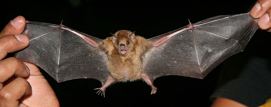
Sturnira lilium (above) and Artibeus planirostris (below), the two bat species in which these new viruses were discovered.
https://upload.wikimedia.org/wikipedia/commons/0/0a/Sturnira_lilium_lostuxtlas2008.jpg
The new viruses are called HL18NL11 and HL17NL10, based on the proteins that make up these viruses. Viruses are typically classified by the structures of their H (hemagglutinin) and N (neuraminidase) proteins. Since the proteins found in these specific viruses don’t carry out the same functions as typical H and N proteins, they are referred to as HL and NL, “hemagglutinin-like” and “neuraminidase-like”, respectively. Both HL17 and HL18 proteins resemble the structure of the H proteins found in conventional influenza A viruses. However, the paper did not investigate the NL proteins very closely, since the function of NL proteins is not well understood.
Although the newly discovered viruses are structurally similar to influenza A viruses, the new viruses have a different mode of transmission from influenza A. Instead of entering the cell by the apical surface, the surface of the cell facing the external environment, they enter the cell by the basolateral surface, the surface that touches neighboring cells—in other words, the cellular equivalent of sneaking in the back door. However, once inside the cell, the viruses are transported to the apical surface to replicate, and the replication and release processes of the new viruses are very similar to those of classic influenza A viruses. Because of their resemblance to these viruses that so often cause disease in birds, the new viruses were tested on cell lines from many different species to see whether they could infect different types of cells.
http://www.pnas.org/content/113/45/12797/F8.large.jpg
Viruses enter by the basal surface of the cells (the surface facing other cells), and new viruses are released from the apical surface (the cell surface facing the external environment).

In the end, most cell lines were resistant to both the original viral genes and the genes injected into VSV (vesicular stomatitis virus, described above). However, they replicated very well in a few cell lines, especially the Madin-Darby canine kidney type II (MDCK II) cell line. More importantly, the viruses were able to infect two human cell lines, which indicates the potential for human infection.

So what does this result mean for us? Most likely, we will not have an HL17NL10 or HL18NL11 outbreak anytime soon. Nevertheless, this research was able to investigate a potential threat. Additionally, the research highlighted the use of an important virological research strategy, the reverse genetic system, for characterizing viruses that won’t grow in lab. Using reverse genetics, the researchers were able to rebuild the viruses using their knowledge of the viral genetic material. In the future, the research team could use this strategy to identify the function of the NL protein in this virus. They could also try injecting both their reverse-genetics-created viral sequences and the VSV-injected viruses into live bats. This would allow them to see if the reverse genetic strategy showed a similar pathology to the original virus, and they could compare the pathogenicity and infectivity of VSV-combined viruses, reverse-genetics-created viruses, and wild-type viruses (the version of the virus that the bats were simply acquiring in the wild). Such experiments would also allow the research team to evaluate the implications of their current findings a little more closely. 

Sources

Note: All sources and pictures are also hyperlinked in text.

    1. Moreira, É. A., Locher, S., Kolesnikova, L., Bolte, H., Aydillo, T., García-Sastre, A., ... & Zimmer, G. (2016). Synthetically derived bat influenza A-like viruses reveal a cell type-but not species-specific tropism. Proceedings of the National Academy of Sciences, 201608821.
    2. Key Facts About Seasonal Flu Vaccine. (2016). Retrieved November 11, 2016, from http://www.cdc.gov/flu/protect/keyfacts.htm
    3. In New Theory, Swine Flu Started in Asia, Not Mexico - The ... (n.d.). Retrieved November 11, 2016, from http://www.nytimes.com/2009/06/24/health/24flu.html
    4.  Main developments in the past 72 hours. (n.d.). Retrieved November 11, 2016, from http://ecdc.europa.eu/en/healthtopics/Documents/100118_Influenza_AH1N1_Situation_Report_0900hrs.pdf 
    5.  Avian influenza. (n.d.). Retrieved November 11, 2016, from http://www.who.int/mediacentre/factsheets/avian_influenza/en/ 

    Photo sources:
    1. https://upload.wikimedia.org/wikipedia/commons/0/0a/Sturnira_lilium_lostuxtlas2008.jpg
    2. https://blogger.googleusercontent.com/img/b/R29vZ2xl/AVvXsEhZYOtDQfA8HbTHStxBiEs36EcGIcSDIuOp4Kx51d-Ea9cKGFFk4iCfQNQJW0xnw4DfPK4StsspXWnOaoYIhBsGoRWu3MQ3YvZikFo7hW17ypYe2ECMJrgXPEwz1Zg76YTHQXIux5wPP-w/s1600/IMG_0620.jpg

    Monday, October 31, 2016

    Dengue Induced Autophagy Through ER Stress: A Mechanism for Viral Replication

    The article reviewed in this post is titled: "Dengue-induced autophagy, virus replication and protection from cell death require ER stress (PERK) pathway activation" by Datan et al 2016. The article was released in March of 2016. 

    Threatening 40% of the world’s population, dengue fever infection has doubled in the past 20 years, (Datan et. al. 2016). A viral disease transmitted to humans by the mosquito genus Aedes, dengue fever involves sudden fever and acute pain in the muscles and joints, (Morens, 2009). Severe dengue fever can turn into dengue hemorrhagic fever and dengue shock syndrome, which can be fatal, (Rajapakse et. al. 2012). As a vector-borne illness, dengue infection varies with seasonal climate changes, but as global warming begins to alter region’s climates, the tropics are no longer the only locations with high risk of dengue. Public health issues associated with the spread of dengue fever make research involving viral mechanisms pertinent to understand how the virus replicates for potential treatments and preventions. In a recent article, investigation of dengue-2 virus reveals that infected cells require ER stress, or PERK, pathway activation for dengue-induced autophagy, viral replication, and protection from other cell stressors, (Datan et. al. 2016). New insight into mechanisms of how dengue replicates can leads to possible treatments and vaccinations as dengue infection advances on the world’s population.
    In order to ensure the virus’s own survival, dengue induces autophagy enabling the viral replication, (Datan et. al. 2016). Previously, dengue-2 was found to kill mice macrophages, yet protect epithelial cells and fibroblasts creating an optimal environment for viral survival. Induced autophagy increased viral replication and viral gene NS4A is upregulated, (McLean et. al. 2011). Inhibition of the NS4A gene limited viral replication indicating the NS4A is sufficient and necessary to trigger PI3K-dependent autophagy, (McLean et. al. 2011).
    Building off this concept, researchers investigated the mechanism of dengue-induced autophagy. Autophagy involved the transport of proteins, lipids, and organelles to autophageosomes, and then they are targeted for lysosomes for degradation as part of a cell’s normal homeostatic response, (Datan et. al. 2016). Typically, the mediators of this process include sensors of cell energy, such as AMPK, however, research has shown dengue virus mediates autophagy through activation of ER, or PERK, pathway activation. Through tracking of calreticulin, a marker of global ER stress, and markers of PERK pathway, ATF-4 (cyclic-AMP-dependent transcription factor 4), and GADD34 (growth arrest and DNA damage-inducible protein 34), ER stress is activated through signaling via the PERK pathway, (Datan et al 2016). Levels of calreticulin, ATF-4, and GADD34 all increased in host cells after infection with dengue virus indicating a strong correlation between viral infection and induced ER stress and PERK pathway signaling, (Figure 1)(Datan et al 2016). Salubrinal, an inhibitor of PERK pathways, eliminates dengue virus induced increases of calreticulin in host cells also strengthening the correlation between ER stress and PERK pathway activation for mechanism of autophagy in dengue-infected cells (Figure 1). The article also described the importance of PERK pathway in viral replication and transcription by treating cells with salubrinal and using specific primers for qPCR. Presence of the PERK pathway inhibitor drastically reduced the transcription of NS4A, the gene previously found to be responsible for autophagy and viral replication in dengue infected cells (Figure 1). Since it is known dengue replication relies on autophagy, the PERK pathway is necessary for viral replication. This information was confirmed with testing in MDCK cells, (Datan et al 2016).
    The study also reviewed how an activated PERK pathway increases authophagy turnover in MEF cells. Using PERK knock-outs and wild-type cells, they measured green puncta of p62, a lysosomal protein and marker for autophagy turnover, via antibodies and observed decreased p62 in PERK wild-types infected with dengue compared to the control, yet not difference in p62 concentrations in PERK knock-outs, (Datan et al, 2016). Since p62 degrades as a result of elevated rates of digestion of autophagosome content in lysosomes, the final step of autophagy, the results from this study further confirm PERK has a necessary role in dengue induced autophagy and thus viral replication.
    The researchers extended the study to determine if ATM (ataxia telangiectasia mutated) signaling followed by the production of ROS (reactive oxygen species), but the results revealed little importance. While direct inhibition of ER stress causes decreases in autophagy turnover, curtails production of ROS and suppresses viral replication, ATM signaling inhibition does little to viral titers and ROS accumulation, (Datan et al 2016). However, they were able to determine that ROS regulates dengue-induced autophagy by implementing a ROS inhibitor linking autophagy to ROS accumulation and then observed high levels of ROS in dengue-infected cells, (Datan et al 2016). Cumulatively, the results demonstrate that dengue virus induces and replicates through autophagy via ER stress through ROS accumulation through a PERK dependent pathway.
    Knowing this mechanism of dengue replication opens up the possibility to treat with pharmaceuticals that interfere with PERK pathway signaling during viral infection. It also poses the possibility that other virus may work this way, providing basis for investigation of other unknown viral mechanisms. For dengue, knowing the mechanisms and inner workings of the virus can help medical professionals and researchers attack the emerging and expanding outbreaks.
    The threat of the reemergence of Dengue Fever also exists for Australia. A 2004 study revealed that intense El Niño Southern Oscillation (ENSO) events combined with surface temperature increases of 0.3-0.6°C correlate with higher incidence rates of mosquitoes-borne illnesses such as Malaria and Dengue Fever, (Zell 2004). Heavy precipitation events lead to floods and development of stagnant water, especially in urban environments, providing viable breeding grounds for mosquito vectors, (Khasnis & Nettleman 2005). Periods of severe rainfall most likely result from increased frequency and intensity of ENSO events due to climate change. Sea level rise creates more extensive saltmarsh habitats along coastlines, providing stagnant, moist environments for mosquitoes, (Russell, 1998). Urban area are also prone to developing more area of stagnant water as heavy rains pool water in tanks, trash bins, gutters and stray rubbish, co-inhabiting locations where mosquitoes reproduce. Increases in temperatures enable geographical shifts of species that carry Dengue Fever, such as Aedes albopictus and Aedes aegypti, further south in Australia, (Russell et. al. 2009). Temperature increases also contribute to quicker virus incubation and elevated rates of vector feeding, demonstrating the potential of rapid transmission to humans.
    A majority of Australia’s population is concentrated in urban areas along the coast. High-density urban areas combined with intense ENSO and QBO events and sea level rise leave the country susceptible to Dengue Fever outbreaks. Present in Australia since the 1880’s, the disease was largely eliminated in the 1950’s, but reappeared in 1981, (Mackenzie et. al. 2006)(Russell, 2009).  Localized around the northern coastlines, dengue fever is expected to move further south and inland, corresponding to climate zone shifts, (Figure 8).  If average surface temperatures in Australia increase by 2.0-3.0°C Dengue Fever could become a common occurrence in Brisbane, (Russell, 2009). Temperature increases of 3.0-4.0°C could stabilize the virus transmission as far south as Sydney. In absence of mitigation strategies to address global warming, the spread of Dengue further south in Australia could occur by 2100 based on warming trends, (Head et. al., 2014).
    Seeing the increased threat of dengue on a global scale, understanding the mechanisms of how the virus replicates initiates better intervention strategies and perhaps prevent the disease from emerging in area affected by climate change. 

    1. Datan, E. Roy, S. G., Germain, G., Zali, N., McLean, J. E., Golshan, G., Harbajan, S. Lockshin, R. A. & Zaken, Z. (2016) Dengue-induced autophagy, virus replication and protection from cell death require ER stress (PERK) pathway activation. Cell Death and Disease. 7: e2127
    2. Head, L., Adams, M., McGregor, H. V. & Toole, S. (2014) Climate Change and Australia. Wiley Interdisciplinary Reviews: Climate Change, 5, 2, 175-197.  
    3. Khasnis, A. & Nettleman, M. (2005). Global Warming and Infectious Disease. Archives of Medical Research, 36, 689-696.
    4. Mackenzie, J., et.al. (2006). Emerging Viral Diseases of Southeast Asia. Issues in Infectious Diseases, 7, 3, 497-504.
    5.  McLean JE, Wudzinska A, Datan E, Quaglino D, & Zakeri Z. (2011) Flavivirus NS4A-inducedautophagy protects cells against death and enhances virus replication. J Biol Chem.  286. 22147–22159.
    6. Morens, David M. MD. Dengue Fever and Dengue Hemorrhagic Fever. Pediatric Infectious Disease Journal. 28. 7. 635-636.
    7. Rajapakse, S., Rodrigo, C. & Rajapakse, A. (2012) Treatment of Dengue Fever. Infection and Drug Resistance. 5. 103-112.
    8. Russell, R. (1998). Mosquito-borne Arboviruses in Australia: The Current Scene and Implications of Climate Change for Human Health. International Journal for Parasitology, 28, 6, 955-969.
    9. Russell, R. (2009). Mosquito-borne Disease and Climate Change in Australia: Time for a Reality Check. Australian Journal of Entomology, 48, 1, 1-7.
    10. Zell, R. (2004). Global Climate Change and the Emergence/Re-emergence of Infectious Diseases. International Journal of Medical Microbiology Supplements, 293, 37, 16-26.

    Sunday, September 25, 2016

    Does Resistance to One Antiviral Indicate Resistance to Another

                Infection by the Hepatitis B virus causes the liver disease Hepatitis B, which can lead to liver failure and cancer.  A simple vaccine can reduce chances of infection, but in places with widespread poverty many people can’t afford the cost of a vaccine.  Subsequently, in such places infection rates remain consistently high leading to an estimated 240 million people worldwide who are chronically infected and 686,000 people who die as a result of Hepatitis B every year.[1]  Due to the epidemic of chronically infected Hepatitis B patients, antiviral drugs have been developed to help prevent the onset of Hepatitis B.  One particular type of antiviral is called a nucleoside analogue.  Such antivirals work by inhibiting viral replication through the use of an alternative DNA subunit, which can be incorporated into viral DNA and subsequently blocks a virus’ own replication machinery.[2]  Nucleoside analogues are exceedingly useful therapies until mutations in the virus’ genetic material causes resistance to the antivirals. 
                A paper[3] published in the journal Nature in July of 2016 by Liu et al. details the usefulness of a specific nucleoside analogue, Entecavir, for treatment of a chronic Hepatitis B infection.  The researchers examined the effectiveness of Entecavir treatment in participants who had never been treated with a nucleoside analogue compared to those who had been treated with one, either lamivudine (LAM) or adefovir dipivoxil (ADV).  The study was conducted over 5 years on 89 individuals from China who were infected with Hepatitis B for at least 6 months and were only treated with Entecavir.  Statistical analysis was used to determine if Entecavir lead to viral remission as associated with prior treatment with LAM, prior history of LAM-resistance, prior history of partial virus resistance to ADV, and prior history of primary treatment failure to ADV. 
                The results of the study indicated that anyone who had never been treated with a nucleoside analogue before exhibited complete viral remission with only a minimal chance of developing resistance.  This is opposed to patients previously treated with a nucleoside analogue, who still responded well to Entecavir but at a lower rate of remission.  The sample group was further characterized by experience with LAM and ADV.  Interestingly, individuals previously treated with LAM, with or without developing resistance, were successfully treated for Hepatitis B, as were ADV patients with partial resistance.  However, ADV-experienced patients who had failed previous treatment had a significantly reduced probability of achieving viral remission when treated with Entecavir. 
                The study by Liu et al. provides important support for reducing antiviral resistance as a result of overuse.  Chronic Hepatitis B can be effectively treated with antivirals, like Entecavir, but for those who failed the first line treatment, doctors should carefully choose the next course of therapy.  By not just prescribing any antiviral when someone has an infection, hopefully the current issue with widespread antibiotic resistance can be avoided, or at least minimized, in regards to antivirals.  The results of the paper strongly support conscientious and judicious use of antiviral drugs.  
                The treatment distinction suggested by Liu et al. for patients with different histories is also indicative of the future of medicine.  Previous means of treating a disease involved a one-size fits all approach.  For example, the paper discusses how ADV was the primary means of treatment for chronic Hepatitis B in China for the past decade.  If a patient was diagnosed with Hepatitis B, he or she was most likely given ADV without deliberation.  However, as the results of the study indicated, such an approach is not effective due to acquired resistance.  Thus, it is important for those in the healthcare profession to consider a patient’s personal history prior to treatment in order to successfully treat an infection, instead of relying on a generalized approach. 
    In order to facilitate the growth of personalized medicine, basic research into the conditions affecting the success of a particular treatment is necessary.  Liu et al. provided important information on a specific drug, but the study was limited by size, as acknowledged by the researchers.  Future research should be more wide spread, meaning more antivirals examined in a larger number of participants with different conditions.  Such work would allow for the development of a comprehensive set of guidelines regarding the use of antivirals.  Analysis of a large set of treatment results could be the beginning of a global initiative for stemming antiviral resistance by providing personalized treatment.


    [1] http://www.who.int/mediacentre/factsheets/fs204/en/
    [2] https://livertox.nlm.nih.gov/NucleosideAnalogues.htm
    [3] Liu, K. et al. A five years study of antiviral effect of entecavir in Chinese chronic hepatitis B patients. Sci. Rep. 6, 28779; doi: 10.1038/srep28779 (2016).

    Zika Virus Causes Problems for Monkeys, Too

    Zika virus is currently a worldwide concern. Spread by mosquitoes and through sexual contact, the virus has been highlighted in the news since May 2015, when it was first detected in Brazil.i Since then, it has taken the world by storm: as of September 22, 2016, it has affected 47 countries/territories in North and South America, with over 275,000 suspected or confirmed cases in Brazil alone.ii (To view the interactive map, please click here.) With all of the fear and news coverage surrounding the outbreak, researchers are racing to learn more about this critical illness. A new paper, published on September 12, 2016, claims to have some of the answers.
    Zika virus infection is often asymptomatic, but for pregnant individuals, the consequences can be devastating. Zika virus is associated with microcephaly (small head) and other fetal malformations.iii In addition, Guillain-Barré Syndrome, a type of autoimmune-induced muscle weakness, has been associated with Zika infection.iii In this research article, Adams Waldorf et al. (2016) primarily investigated the virus’s effects on fetal brain development. For this experiment, the team injected a Cambodian strain of the virus (strain FSS13025, Cambodia 2010) into five locations on the forearms of a pregnant pigtailed macaque monkey (see image of pigtailed macaque below). The monkey was had been pregnant for 119 days, approximately equivalent to 28 weeks of human pregnancy. The fetus was viewed by ultrasound weekly, and cesarean section was performed 43 days later (equivalent to 38 weeks in human pregnancy). The results were simply astonishing.


    Within 10 days of the virus injection, the developing fetus began to encounter problems. The fetus showed lesions (damaged areas) on its brain, which evolved differently on the left and right hemispheres. In the left hemisphere, the fetus showed loss of brain volume, and even ventricular collapse (complete destruction of a ventricle, a hollow area within the brain). In the right hemisphere, the lesions simply increased in severity over time. White matter, one of the types of brain tissue consisting mainly of nerve fibers, stopped growing over time, while gray matter, another type of brain tissue consisting mainly of nerve cell bodies, continued to grow. The picture below shows some of this damage, indicated by red arrows (the numbers at the top signify the number of days after injection/the number of days since conception).

    After cesarean section, the fetus was autopsied for further investigation. Evidence of the virus was detected in the brain, which showed significant underdevelopment of white matter. Brain lesions were observed throughout. These findings are very similar to reports of Zika virus in human fetuses, as seen by magnetic resonance imaging (MRI) after birth.
    As groundbreaking as this study is, there are several flaws in its design. Only a single subject was investigated, and this subject was a pigtail macaque monkey. Meanwhile, a previous study conducted on rhesus macaques failed to produce similar results.iv  Therefore, the result of this pigtail macaque study could be coincidental or unrepresentative of typical Zika effects. Also, this study used five injection sites, giving a very high dose of the virus, which may not be reflective of an ordinary bite from a virus-carrying mosquito.
    Future studies should include larger sample sizes, and should include several different types of macaque monkeys or similar species. In addition, future research could explore the mechanism causing the observed symptoms. How does the virus cause damage to the developing fetus? Lastly, more research needs to be conducted to determine if the pigtailed macaque could be a good model on which to test new medications for Zika virus. If pigtailed macaque monkeys respond very similarly to humans, then perhaps potential new vaccines could be tested on them, before the vaccines move to human trials.


    Paper: Waldorf, K. M. A., Stencel-Baerenwald, J. E., Kapur, R. P., Studholme, C., Boldenow, E., Vornhagen, J., ... & Armistead, B. (2016). Fetal brain lesions after subcutaneous inoculation of Zika virus in a pregnant nonhuman primate. Nature Medicine.

    Other sources:
    iBBC News (August 31, 2016). Zika outbreak: What you need to know. Retrieved September 25, 2016, from http://www.bbc.com/news/health-35370848
    iiEpidemic Diseases - Zika in the Americas. (September 22, 2016). Retrieved September 25, 2016, from http://ais.paho.org/phip/viz/ed_zika_countrymap.asp
    iiiWorld Health Organization. 2016. Zika virus, microcephaly and Guillain-Barré syndrome. World Health Organization, Geneva, Switzerland. Retrieved September 25, 2016 from http://apps.who.int/iris/bitstream/10665/204961/1/zikasitrep_7Apr2016_eng.pdf?ua=1
    ivDudley, D. M., Aliota, M. T., Mohr, E. L., Weiler, A. M., Lehrer-Brey, G., Weisgrau, K. L., ... & Gellerup, D. D. (2016). A rhesus macaque model of Asian-lineage Zika virus infection. Nature communications, 7.