Zika virus is currently a worldwide
concern. Spread by mosquitoes and through sexual contact, the virus has
been highlighted in the news since May 2015, when it was first detected in
Brazil.i
Since then, it has taken the world by storm: as of September 22, 2016, it has
affected 47 countries/territories in North and South America, with over 275,000
suspected or confirmed cases in Brazil alone.ii
(To view the interactive map, please click here.) With all
of the fear and news coverage surrounding the outbreak, researchers are racing
to learn more about this critical illness. A new paper, published on September
12, 2016, claims to have some of the answers.
Zika virus infection is often
asymptomatic, but for pregnant individuals, the consequences can be
devastating. Zika virus is associated with microcephaly (small head) and other fetal
malformations.iii
In addition, Guillain-Barré Syndrome, a type of autoimmune-induced muscle
weakness, has been associated with Zika infection.iii In this
research article, Adams Waldorf et al.
(2016) primarily investigated the virus’s effects on fetal brain development.
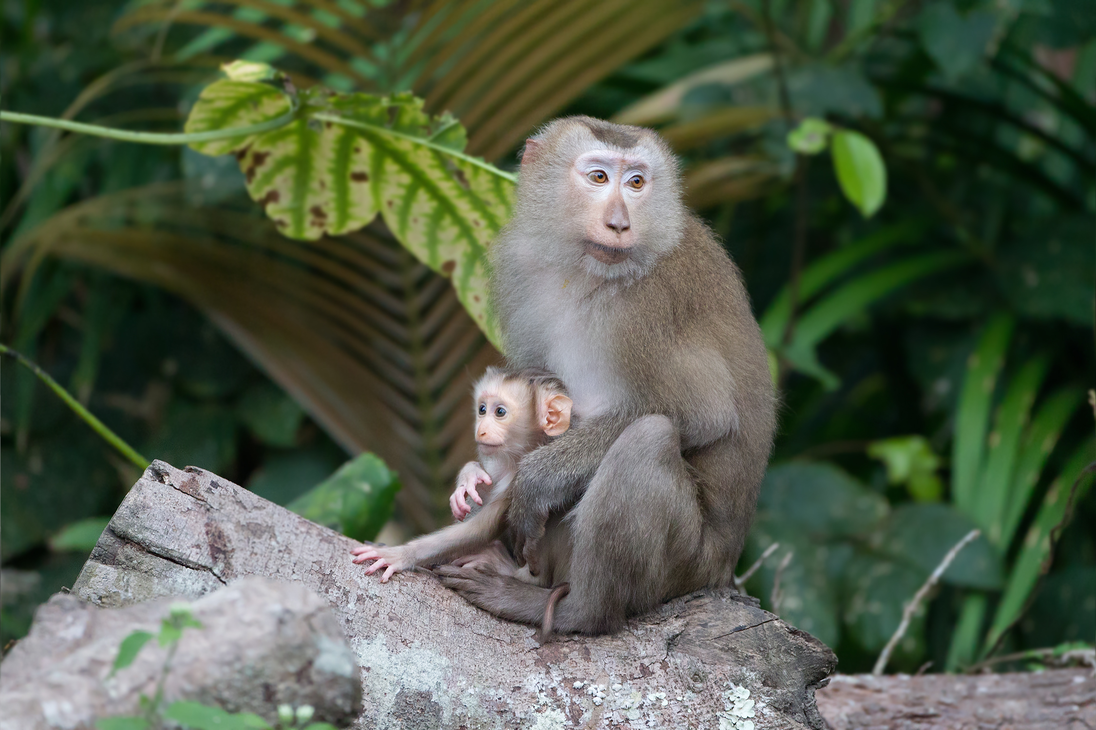
For this experiment, the team injected a Cambodian strain of the virus (strain
FSS13025, Cambodia 2010) into five locations on the forearms of a pregnant
pigtailed macaque monkey (see image of pigtailed macaque below). The monkey was had been pregnant
for 119 days, approximately equivalent to 28 weeks of human pregnancy. The
fetus was viewed by ultrasound weekly, and cesarean section was performed 43
days later (equivalent to 38 weeks in human pregnancy). The results were simply
astonishing.
Within 10 days of the virus
injection, the developing fetus began to encounter problems. The fetus showed
lesions (damaged areas) on its brain, which evolved differently on the left and right
hemispheres. In the left hemisphere, the fetus showed loss of brain volume, and
even ventricular collapse (complete destruction of a ventricle, a hollow area
within the brain). In the right hemisphere, the lesions simply increased in
severity over time. White matter, one of the types of brain tissue consisting
mainly of nerve fibers, stopped growing over time, while gray
matter, another type of brain tissue consisting mainly of nerve cell bodies, continued to grow. The picture below shows some of this damage, indicated by red arrows (the numbers at the top signify the number of days after injection/the number of days since conception).

After cesarean section, the fetus
was autopsied for further investigation. Evidence of the virus was detected in
the brain, which showed significant underdevelopment of white matter. Brain
lesions were observed throughout. These findings are very similar to reports of
Zika virus in human fetuses, as seen by magnetic resonance imaging (MRI) after
birth.
As groundbreaking as this study is,
there are several flaws in its design. Only a single subject was investigated,
and this subject was a pigtail macaque monkey. Meanwhile, a previous study
conducted on rhesus macaques failed to produce similar results.iv Therefore, the result of this pigtail macaque
study could be coincidental or unrepresentative of typical Zika effects. Also,
this study used five injection sites, giving a very high dose of the virus,
which may not be reflective of an ordinary bite from a virus-carrying mosquito.
Future studies should include
larger sample sizes, and should include several different types of macaque
monkeys or similar species. In addition, future research could explore the
mechanism causing the observed symptoms. How does the virus cause damage to the
developing fetus? Lastly, more research needs to be conducted to determine if the
pigtailed macaque could be a good model on which to test new medications for
Zika virus. If pigtailed macaque monkeys respond very similarly to humans, then
perhaps potential new vaccines could be tested on them, before the vaccines
move to human trials.
Paper: Waldorf, K. M. A., Stencel-Baerenwald, J. E., Kapur, R. P., Studholme, C., Boldenow, E., Vornhagen, J., ... & Armistead, B. (2016). Fetal brain lesions after subcutaneous inoculation of Zika virus in a pregnant nonhuman primate. Nature Medicine.
Image source: https://upload.wikimedia.org/wikipedia/commons/9/96/Macaca_leonina_mother_with_baby_-_Khao_Yai.jpg
Other sources:
iBBC
News (August 31, 2016). Zika outbreak: What you need to know. Retrieved
September 25, 2016, from http://www.bbc.com/news/health-35370848
iiEpidemic
Diseases - Zika in the Americas. (September 22, 2016). Retrieved September 25,
2016, from http://ais.paho.org/phip/viz/ed_zika_countrymap.asp
iiiWorld Health Organization. 2016. Zika virus,
microcephaly and Guillain-Barré syndrome. World Health Organization, Geneva,
Switzerland. Retrieved September 25, 2016 from http://apps.who.int/iris/bitstream/10665/204961/1/zikasitrep_7Apr2016_eng.pdf?ua=1
ivDudley,
D. M., Aliota, M. T., Mohr, E. L., Weiler, A. M., Lehrer-Brey, G., Weisgrau, K.
L., ... & Gellerup, D. D. (2016). A rhesus macaque model of Asian-lineage
Zika virus infection. Nature
communications, 7.
No comments:
Post a Comment