Novel Role of p53 in Septic Immunosuppression: Involvement in Loss and Dysfunction of CD4+ Lymphocytes
published in November, 2018 by Zhang et al.
Approximately half of human cancers express mutations in the gene for p53, an important transcription
factor that regulates cell proliferation and apoptosis [1]. Therefore, p53 is usually investigated and
discussed in the context of tumors. While its role in controlling tumorigenesis has been thoroughly
researched, few studies have examined the role of p53 in immune cells. In this paper, Zhang et al. demonstrate how p53 can also function to suppress proliferation of CD4+
lymphocytes and initiate apoptosis, resulting in septic immunosuppression [2]. The diagram below
depicts the role of p53 in the intrinsic apoptotic pathway (Figure 1).

Figure 1. p53 upregulates pro-apoptotic proteins Bax and Apaf-1, resulting in the formation of the apoptosome and
the induction of the apoptotic cascade.
the induction of the apoptotic cascade.
Sepsis is the body’s response to an extreme infection, which typically results in immunosuppression [3,4].This study demonstrated that as a result of sepsis in mice, levels of p53 were elevated in CD4+ T
lymphocytes. This p53 upregulation likely inhibited T cell proliferation while inducing apoptosis and
immune dysfunction in T cells.
lymphocytes. This p53 upregulation likely inhibited T cell proliferation while inducing apoptosis and
immune dysfunction in T cells.
In order to determine the effects of sepsis on CD4+ lymphocytes, the authors conducted a series of
experiments. First, they produced sepsis in C57BL/6 mice via cecal ligation and puncture (CLP) to see if sepsis in mice would cause loss and dysfunction of CD4+ lymphocytes [5].
The results demonstrated that CD4+ lymphocytes isolated from these mice lacked effective proliferative
abilities (Figure 2). Furthermore, the CD4+ lymphocytes gradually underwent apoptosis after CLP. There
was also a significantly lower level of IL-2 production in septic mice, indicating a decline in T cell
proliferation, as well as an increase in the IL-4/IFN-γ ratio in mice after CLP, indicating a shift to an immunosuppressive Th2 response (Figure 3) [2].
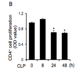
Figure 2. CD4+ T cell proliferation significantly decreases 24 and 48 hours after CLP treatment.
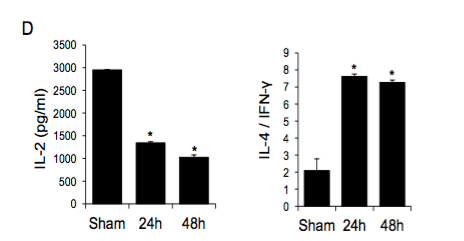
Figure 3. IL-2 production in CD4+ T cells decreases after CLP treatment and IL-4/IFN-γ ratio increases after CLP treatment, indicating a shift to the immunosuppressive Th2 response.
After the authors determined that CLP in mice resulted in loss and dysfunction of CD4+ T lymphocytes,
they demonstrated that there was enhanced p53 expression in CD4+ T lymphocytes after sepsis. By
measuring protein and mRNA expression levels using western blotting and real-time PCR, the authors
showed that p53 mRNA was upregulated in splenic CD4+ T lymphocytes of mice after CLP (Figure 4).
they demonstrated that there was enhanced p53 expression in CD4+ T lymphocytes after sepsis. By
measuring protein and mRNA expression levels using western blotting and real-time PCR, the authors
showed that p53 mRNA was upregulated in splenic CD4+ T lymphocytes of mice after CLP (Figure 4).
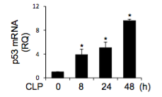
Figure 4. p53 expression is significantly upregulated in CD4+ T lymphocytes 8, 24, and 48 hours after CLP treatment in mice.
In order to determine the function of p53, the authors then blocked its activity by injecting its inhibitor, PFT, prior to CLP treatment [6]. In the presence of PFT, the mortality rate of the mice decreased. This
finding suggests that p53 was involved in the mortality of septic mice. The authors compared CD4+ T cell
proliferation in mice treated with CLP alone to that of mice treated with PFT and CLP. The results
demonstrated that when p53 was left uninhibited, it resulted in significantly less CD4+ T cell proliferation than when p53 was inhibited by PFT (Figure 5). This finding suggests that p53 plays a role in suppressing CD4+ T cell proliferation.
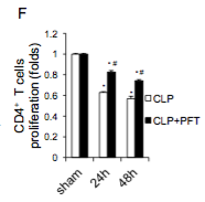
Figure 5. p53 plays a role in inhibiting CD4+ T cell proliferation. CLP induced inhibition of proliferation was diminished by PFT injection. In other words, when p53 was inhibited, CD4+ T cells were able to proliferate.
Next, the authors demonstrated how p53 mediates apoptosis of CD4+ T lymphocytes during sepsis.
LPS stimulation in vitro was also used to mimic a septic environment in the mice. Similarly to CLP,
LPS treatment induced upregulation of p53, resulting in decreased CD4+ T cell proliferation. Furthermore,
when treated with LPS, CD4+ T lymphocytes had an elevated level of Bax, a pro-apoptotic regulator [7].
LPS treatment also initiated caspase-3 cleavage, which is the first step of the apoptotic cascade (Figure 6)
[8]. Caspase-3 cleavage was blocked by p53 inhibition, demonstrating that p53 is necessary for mediating
apoptosis of CD4+ T lymphocytes.

Figure 6. A schematic diagram of the apoptotic pathways. The intrinsic pathway on the left is relevant to this paper, as this diagram represents the roles of Bax and caspase-3 in the apoptotic pathway. Bax pokes holes in the mitochondrial membrane. The subsequent release of cytochrome C leads to the cleavage of caspase-3, which induces the apoptotic cascade. When treated with LPS, CD4+ T lymphocytes had an elevated level of Bax, and LPS initiated caspase-3 cleavage.
As previously stated and demonstrated in Figure 3, inactivated T cells in septic environments with p53
had a decrease in IL-2 production and an increase in IL-4/IFN-γ ratio. These cytokine differences are important because they tell us that the T cells became inactivated. The binding of IL-2 to CD25 is required for T cell activation, so less IL-2 production means less T cell activation. Furthermore, an increased IL-4/IFN-γ ratio means that a septic environment resulted in a higher presence of IL-4, which functions to differentiate Th0 cells into Th2 cells. Th2 cells are immunosuppressive, and therefore, they tamp down the immune response. These cytokine phenotypes were almost normal when treated with p53 inhibitor, suggesting that inactivation of these CD4+ T lymphocytes involved p53. Also, in p53 knockout mice, sepsis did not suppress IL-2 production, suggesting that p53 serves a role in suppressing IL-2 production and thus CD4+ T cell proliferation [2].
had a decrease in IL-2 production and an increase in IL-4/IFN-γ ratio. These cytokine differences are important because they tell us that the T cells became inactivated. The binding of IL-2 to CD25 is required for T cell activation, so less IL-2 production means less T cell activation. Furthermore, an increased IL-4/IFN-γ ratio means that a septic environment resulted in a higher presence of IL-4, which functions to differentiate Th0 cells into Th2 cells. Th2 cells are immunosuppressive, and therefore, they tamp down the immune response. These cytokine phenotypes were almost normal when treated with p53 inhibitor, suggesting that inactivation of these CD4+ T lymphocytes involved p53. Also, in p53 knockout mice, sepsis did not suppress IL-2 production, suggesting that p53 serves a role in suppressing IL-2 production and thus CD4+ T cell proliferation [2].
So why does this matter?
While there were various research questions posed and experiments conducted, the main takeaway from
this paper is that in septic CD4+ T lymphocytes, expression of p53 is upregulated. As a result of this
upregulation, proliferation of these CD4+ T cells is inhibited, and apoptosis is induced. A decreased level
of IL-2 is secreted, tamping town proliferation of T cells, while an increased level of IL-4 is secreted,
shifting the T cell to a Th2 immunosuppressive response. The findings of this paper are significant
because they demonstrate a new role for p53. While most of the previously published literature on p53
is about its role as a tumor suppressor that helps regulate the growth of tumors, this paper sheds light
on a potentially detrimental role of p53 in inducing loss and dysfunction of CD4+ T lymphocytes in
septic environments. p53 is typically known for its ability to induce apoptosis in tumor cells, which is
beneficial to our health. However, this study demonstrates how p53 can also induce apoptosis in CD4+
T cells and diminish their immune function, which can be detrimental to our health. Thus, it is important
to remember that genes can have specific roles in different contexts. There are still many mechanisms
involved in the activation of p53 in immune cells that remain unclear. Therefore, in future experiments,
it would be interesting to further investigate what specific mechanism causes upregulation of p53 in
eptic cells. It may be worth further investigating the relationship between p53 upregulation and
production of specific cytokines. For example, what specific mechanism causes a decrease in IL-2
production? Similarly, what specific mechanism causes an increase in IL-4 production? How is p53
involved in these mechanisms? Finally, it would be interesting to see if these results are similar in septic
patients or if they are unique to these C57BL/6 mice. The application of these findings to the medical
field could be extremely beneficial for beginning to understand how to combat immune suppression
induced by sepsis.
Works Cited
[1] Vogelstein, Bert et al. "p53: The Most Frequently Altered Gene in Human Cancers." Nature Education 3 (2010). https://www.nature.com/scitable/topicpage/p53-the-most-frequently-altered-gene-in-14192717.
[2] Zhang, H. et al. "Novel Role of p53 in Septic Immunosuppression: Involvement in Loss and
Dysfunction of CD4+ Lymphocytes." Cell Physiol Biochem (2018). 452 - 469.
Dysfunction of CD4+ Lymphocytes." Cell Physiol Biochem (2018). 452 - 469.
[3] "What is sepsis." Centers for Disease Control and Prevention, 20 July 2018. https://www.cdc.gov/sepsis/what-is-sepsis.html.
[4] "NCI Dictionary of Cancer Terms." National Cancer Institute, https://www.cancer.gov/publications/dictionaries/cancer-terms/def/immunosuppression.
[5] Toscano, M. G. et al. "Cecal ligation puncture procedure." J Vis Exp (2011). https://www.ncbi.nlm.nih.gov/pubmed/21587163.
[6] "Pifithrin-alpha." Sigma-Aldrich, Sigma-Aldrich Inc., https://www.sigmaaldrich.com/catalog/product/sigma/p4359?lang=en®ion=US. [7] "UnitProKB - Q07813 (BAX_MOUSE)." UniPro, UniPro Consortium, 2018. https://www.uniprot.org/uniprot/Q07813.
[8] "UnitProKB - P42574 (CASP3_HUMAN). UniPro, UniPro Consortium, 2018. https://www.uniprot.org/uniprot/P42574.
No comments:
Post a Comment