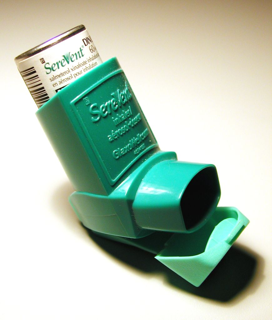
Asthma is a “type I hypersensitivity reaction,” meaning that it is caused by the secretion of a
particular type of antibody, IgE, following a Th2 immune response. Th2
responses are one of the major types of immunity, and target extracellular
pathogens. The Th2 class of helper T cells secrete cytokines that stimulate B
cells to produce IgE, which can activate other types of cells to respond to
antigens. When these IgE antibodies are targeted against environmental
allergens, and not pathogens, type I hypersensitivity results, and when the
response occurs in the bronchioles, it results in asthma. A recent study at the
Mayo Clinic Arizona on asthma examined the role of eosinophils, a type of
specialized white blood cell, in the regulation of dendritic cells and Th2 pulmonary
immune responses with T cells in asthmatic mice. There have been rumblings that the current description of
eosinophils as “end-stage destructive effector cells” is not broad enough and
that they in fact are involved with the secondary immune responses which lead
to the activation and proliferation of memory T cells. The DCs present the antigen to the
lungs; in studies of asthma in mice, including this one, the mice were given
additional DCs to better spread the allergen triggering antigen. If a mouse has fewer eosinophils, then
they generally will have reduced Th2 pulmonary abilities. This study suggests that in allergen-specific T cell
responses, eosinophils and DCs work together and are equally important and lead
to a Th2 polarized immune response.
The eosinophils were found to have a profound impact on whether the
inflammation of the respiratory tract goes by a Th2 pathway or a Th17 (a
different “flavor” of immune response) pathway.
The relationship
between the location of the eosinophils and their abundance, as compared with
the accumulation of T cells during an asthma attack, was studied with three
types of mice: eosinophil-null (PHIL mice), eosinophil-sufficient (wild type)
and eosinophil-low (IL-5-/- mice). PHIL mice had absolutely no eosinophils in their system,
while the IL-5-/- mice had a partial loss of eosinophils compared to
the wild type. The mice were all
treated with injections of an innocuous allergen under conditions that trigger
asthma (OVA/Alum) and were subsequently exposed to airborne antigens. The effects of the airborne antigens on
the mice were then examined. The
comparisons between the three types of mice reveal information about whether or
not the mice were able to accumulate DCs and eosinophils. The draining lymph nodes of the PHIL
mice did not acquire DCs in the 20 hours after they were exposed to the airborne
antigens. In contrast, within 20 hours the nodes of the wild type mice acquired
both eosinophils and MHC II activated DCs. The researchers concluded that eosinophils are necessary for
DCs to move to the lymph nodes and to cause T cell activation. The mice that lacked the proper amount
of eosinophils could not have the correct Th2 polarization in their lungs once
they were exposed to an antigen, as the T cells were not properly activated in
the LDLNs.
There is certainly
more to be learned about eosinophils, DCs, T cells and their role in causing
asthma. The researchers suggest
that eosinophils as monitors of localized immune responses can provide
wonderful insights into the wider role of eosinophils in the immune system, as
their impact on inflammation must certainly involve more than just allergies
and asthma.
Jacobsen, E. A., Zellner, K. R., Colbert, D., Lee, N. A.,
& Lee, J. J. (2011). Eosinophils regulate dendritic cells and Th2 pulmonary immune responses following allergen provocation. The Journal of Immunology,
187, 6059-6068.
Post by Jessie Solcz
Post by Jessie Solcz

Its a nice blog informing about asthma medications
ReplyDelete